The prognosis of a skin cancer can be very different depending on how long it has been growing on our skin. One of the great challenges in dermatology is to detect malignant lesions as early as possible. For this we have a technology called digital dermoscopy or epiluminescence microscopy, which is one of the most modern and accurate methods for detecting changes in skin lesions.
As a dermatologist, from the melanoma unit of the Hospital Clínic de Barcelona.I have seen how patients who are in a digital mole tracker can make very early diagnoses of malignant lesions and thus perform curative treatments.
If you have been referred for this test and you are looking for a you can consult this link for more information. If you also want to know what this test is all about, you can continue reading this blog where I explain in depth what digital dermoscopy is all about and its benefits.
I made this article as graphic as possible and with many images so that it can be perfectly understood.
What will I talk about in this article?
Evolution of mole (nevus) examination over the years.
Medicine has evolved rapidly over the last few years and dermatology has always been a pioneer in incorporating new imaging techniques and recently artificial intelligence into clinical practice. Likewise, the way of assessing the skin has also been evolving rapidly over the last decades.
In general, many years ago we used only our clinical eye to look for changes in moles or suspicious lesions. However, in this way, we were not able to detect small changes in moles to find skin cancer at early stages.
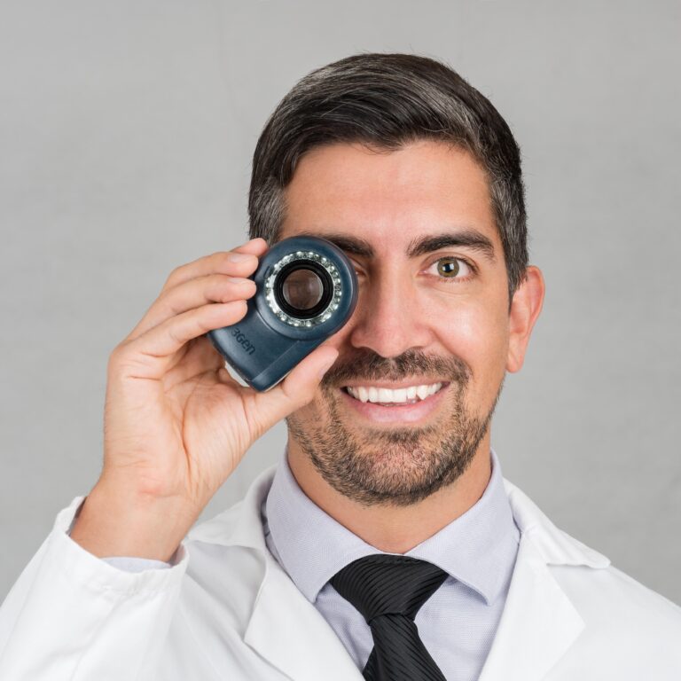
Subsequently, more than 20 years ago, dermatoscopy was introduced in our practicesThe magnifying glass, which is a high magnification magnifying glass with polarized light that allows us to see through the skin. In this way we can evaluate new structures that are not possible with the naked eye. In this way we were able to increase our diagnostic accuracy tremendously.
And, since the beginning of the year 2000, we started to digitally track moles. With this system we are able to digitally record body maps (photos of the whole body) and also dermatoscopic images of moles and follow them over time. With this method, applied in patients at risk of developing skin cancer, we have been able to significantly improve the early detection of skin cancer.
What is digital dermoscopy?
Digital dermatoscopy or epiluminescence microscopy is a dermatological test that allows us to track moles very accurately. The digital dermoscopy technique incorporates an image archiving system that allows comparison of all subsequent examinations performed.
Thanks to these characteristics, it is easier to detect and observe the slightest changes, either in shape or color, of the pigment.
For whom is digital dermoscopy indicated?
Patient follow-up with digital dermoscopy is indicated in those patients who have risk factors for developing melanoma. Among them are:
- Patients with many moles
- Patients with clinically atypical moles. See the ABCDE rule in the following link.
- Risk phototypes such as having very light skin with a tendency to sunburn.
- Patients with a history of melanoma
- Family history of melanoma
- Genetic mutations that predispose us to melanoma. We perform mutational studies in patients who have more than 2 melanomas and at least one of them before the age of 60. Also in patients who have had 1 melanoma and a first-degree relative with 1 melanoma before the age of 60.
- Having used tanning booths or having had significant sun exposure.
- Patients with immunosuppression, such as solid organ transplant recipients requiring immunosuppressants.
How is the visit including digital dermoscopy performed?
Currently, the most accepted procedure to perform a digital follow-up has 2 stages, first we will perform a total body mapping and a dermatoscopic evaluation of the suspicious moles.
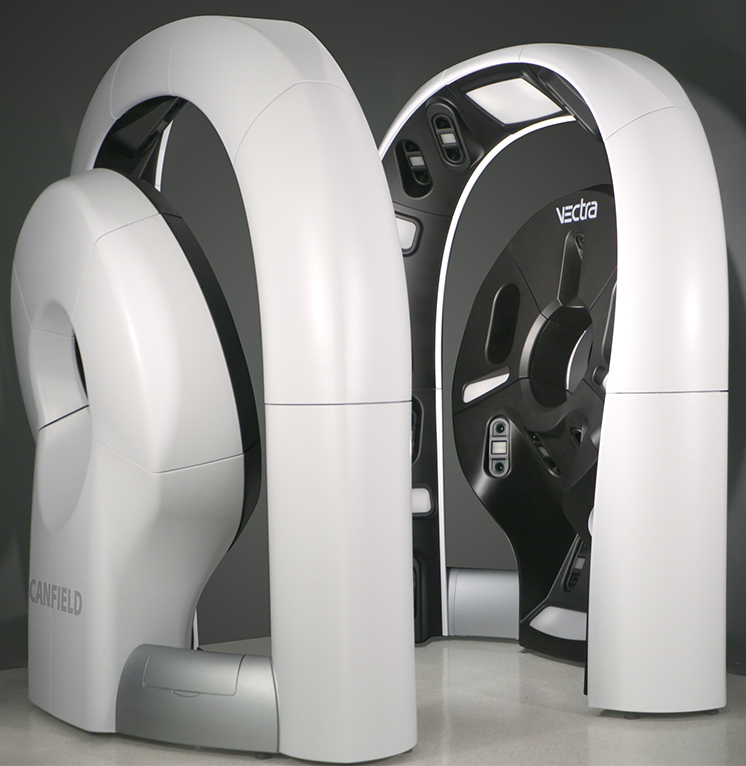
For many years we have had 2D tracking systems but recently, and only in a few places in Spain (3 to be precise), we have 3D technology.
However, for safety reasons, we always do some additional steps, which I will tell you about below:
Stage 1
Body maps in 2 or 3 dimensions. First of all, we will take some complete body photographs in order to check if there are any general changes between the previous and the current follow-ups.
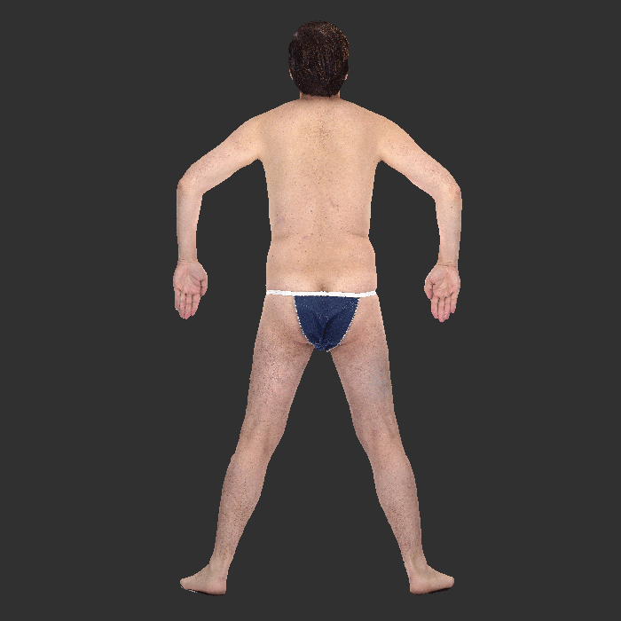
In this animation you can see how your body will be reconstructed after having taken the body photos using a 3D system. The great advantage of this technology is that it takes only seconds to take the photos.
Stage 2
Digital dermoscopy. Secondly, we will perform a microscopic examination of the entire skin surface with dermoscopy, locating the most atypical lesions, photographing them for subsequent follow-up.
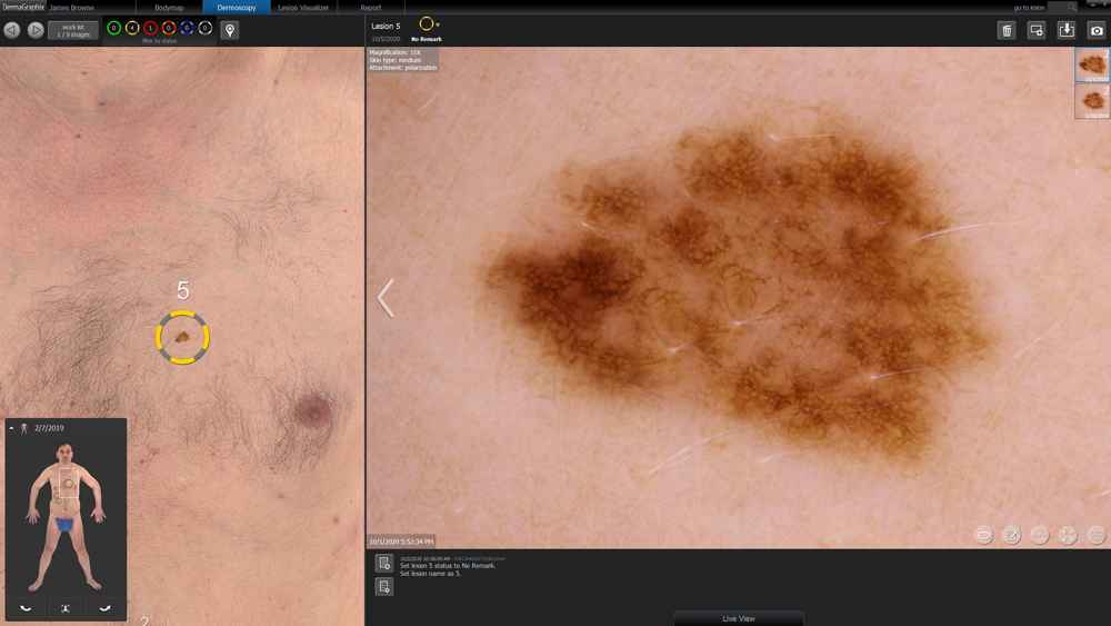
In this image you see how we located a slightly more atypical mole on the left and then recorded a high-resolution dermoscopic image for future follow-up.
Stage 3: complete dermatological check-up
Manual examination with dermoscopy of the entire skin. Although we have a lot of technology we will never replace the expertise of a dermatologist for the follow-up of your moles. That is why the The last step is the most important, where we always perform a manual physical examination with our dermatoscope. to evaluate all the lesions or moles you have on your body.
This step is very important because it allows us to check moles that we have not recorded in the digital dermoscopy and to check "hidden" sites that are more difficult to follow with photos, such as the scalp, behind the ears or between the fingers.
Also, we will ask if you have noticed any "spots" or moles in the genital area or also in the mucosa of the mouth because these are places where melanoma can also appear.
How do we evaluate changes in digital dermoscopic monitoring?
Depending on the risk of having a melanoma in the future, we will decide how often you need to come for check-ups. Normally it will be between 6 to 12 months.
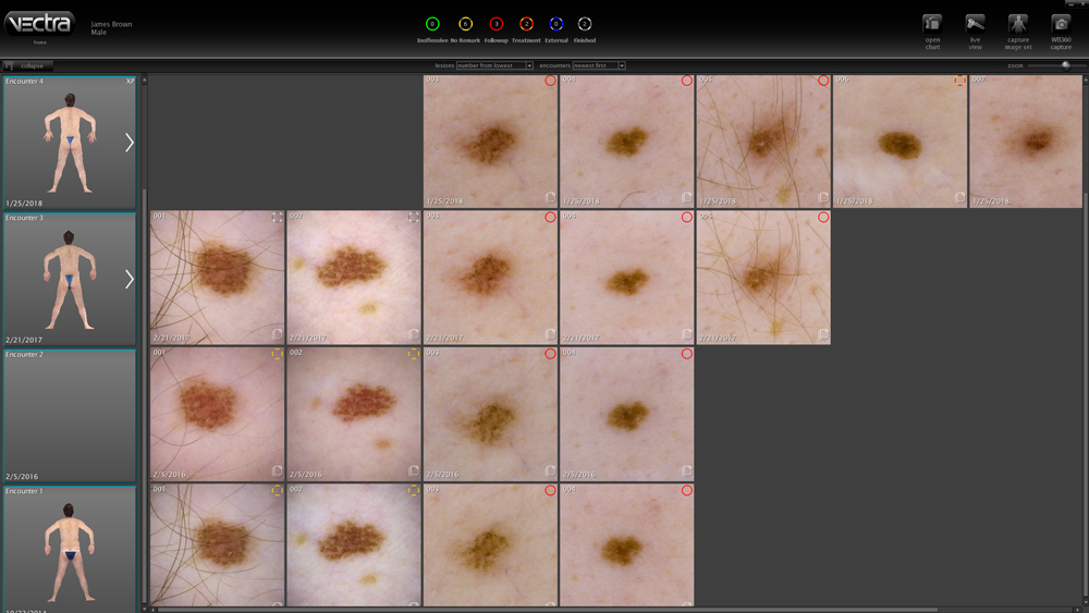
In this photograph we can see the digital dermoscopic follow-up of a patient who has come 4 times to the office. From bottom to top we can see the individual evolution of each mole. In this case we do not observe any important change so the patient can be very calm.
Benefits of performing digital dermoscopy for mole monitoring
There are several benefits in selected patients (see section, for whom is it indicated?) to perform this technique.
- Allows the early detection of skin cancer of melanoma and non-melanoma type
- Benefit of not overlook melanoma with few dermatoscopic criteria.
- Allows improve prognosis of patients.
- Reduce the number of unnecessary excisions or biopsies.
Is there scientific evidence that digital dermoscopy is useful?
- Multiple cohort studies consistently show that digital dermoscopy allows the detection of very early melanomas that are not yet detected. lack clinical or dermoscopic evidence of malignancy. [1] [2] [3] [4] [5] [6]
- In addition, multiple studies have shown that the impact of the routine use of digital dermoscopy is high in terms of the proportion of melanomas detected by the technique. In three studies (two prospective observational clinical trials and two prospective [7] [8] and a retrospective cohort [9] of moderate-high risk patients in a specialized setting, epilimuniscence microscopy was able to detect between 34 and 61% of the patients' melanomas.
- Finally, in two prospective observational trials at the hospital level,[10] and another in primary care,[11] demonstrated that digital dermatoscopy significantly reduces the proportion of excisions of benign lesions:melanomas. In other words, by performing digital dermoscopy we can avoid multiple mole removals that are not necessary.
Where can you follow up your moles with digital dermoscopy?
If you would like to request a digital mole follow-up visit in Barcelona, SpainYou can click on the following link to request an appointment with me.
In dermatological diagnosis we specifically count with the VECTRA WB360 full-body 3D imaging system, which in Catalonia is only available at the Hospital Clínic de Barcelona and the Diagnosis dermatológica clinic. The advantage of this equipment is that it captures the entire surface of the skin with macroscopic quality resolution in just seconds and has several artificial intelligence tools to improve the detection of changes over time.