Skin cancer
Rely on a dermatologist with skin cancer expertise for peace of mind
Tell me about your problem

Why am I an expert dermatologist in skin cancer?
Patients
I visit more than 1,000 patients with skin cancer or at high risk of developing it every year.
Research
Active researcher in the field of skin cancer with more than 60 high impact articles in international journals.
Teaching
I am the teaching coordinator of the Melanoma Unit at Hospital Clínic de Barcelona.
First visit
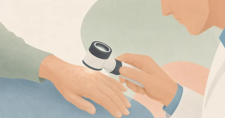
Often the least expected nevi (moles) can be the ones that are most likely to be and can evolve into skin cancer. That is why during your first visit with a dermatologist in skin cancer we will perform a individualized valuation of your nevus and also of the risk factors you may have. In the first visit we will always include dermatoscopy that will allow me to identify minimal changes in the nevi that may be present. may suggest malignancy.
1
Background
We will assess your personal and family history of risk for developing skin cancer.
2
Dermatoscopy
In a second step we will evaluate all moles on your body by manual dermoscopy.
3
Follow-up
Based on your individual risk we will decide if you need to perform specific monitoring using digital monitoring equipment.
Risk factors for developing skin cancer
Knowing the risk factors for developing skin cancer is important for early detection strategies for skin cancer. A dermatologist who is an expert in skin cancer is able to detect changes in your moles early and offer you immediate treatment. There are many risk factors that can increase the risk of developing skin cancer but here are the most common ones.
Sun exposure
Ultraviolet radiation from the sun irreversibly damages the genes of skin cells. Tanning booths are even worse as they provide twice as much radiation as the sun.
Phenotype
There are certain phenotypes of higher risk such as having many moles, blond-red hair, having many freckles on the face or having light-colored eyes.
Family history
Melanoma is a highly heritable cancer. Therefore, having a family history of melanoma increases the risk of developing it.
Personal background
Approximately 5% to 10% of people with melanoma will develop a new melanoma in their lifetime.
What is dermoscopy?

La dermatoscopy is a non-invasive technique used by dermatologists who are experts in skin cancer. A dermatoscope has polarized light and a powerful magnifying lens that allows us to visualize structures through the skin that are normally invisible to the naked eye.
Thanks to dermatoscopy, we can make a diagnosis with greater precisionThis reduces the need for invasive procedures, such as biopsies or excision. In your case as a patient, this reduces the possibility of unsightly scarring or the stress of surgery.
How do my nevi look with dermoscopy?

I have a lot of moles! What can I do?
If you have many moles and some of them from atypical characteristicsor if you have risk background for the development of melanoma, you may be a candidate for specific monitoring. A good option will be the use of specialized digital tracking. Thanks to this modern equipment, which includes the use of artificial intelligence, we will be able to detect changes in the nevi at a very early stage. These millimeter changes will allow us to identify suspicious lesions very early and offer curative treatment. Below you can see how the digital follow-up of a patient is performed using a modern imaging system called photofinder.
Moles difficult to visualize
Confocal microscopy

On some occasions there may be nevi that are very difficult to diagnose clinically or in delicate areas where it would be preferable not to perform a biopsy, such as the face. This is why we have confocal microscopy equipment that allows us to perform biopsies in a very accurate way. non-invasive obtain microscopic resolution images and be able to make a diagnosis of certainty, without having to remove a mole.
Here we can see an image of confocal microscopy of a suspicious lesion. This cellular resolution image allowed us to make the diagnosis of certainty of a melanoma that could be removed early and the patient was cured.

I have skin cancer and need treatment
There are various types of skin cancer that can affect us but the main ones are basal cell carcinoma, squamous cell carcinoma and melanoma. It is important to know that not all skin cancers are the same and that each patient is unique. During our visit we will assess the diagnosis of your skin cancer and seek to offer you a treatment with curative intent.
Do you want to start taking care of yourself and prevent skin cancer with a dermatologist who is an expert in skin cancer?
See my blog

Squamous cell carcinoma: symptoms, risk, and treatment
Squamous cell carcinoma is the second most common skin cancer and also one of those with the best prognosis when detected early. I will explain to you, without alarmism, how it develops from a precancerous lesion, what makes it more or less aggressive, how to recognize it, and what treatment corresponds to each case.
Field cancerization of the face: what it is and treatment
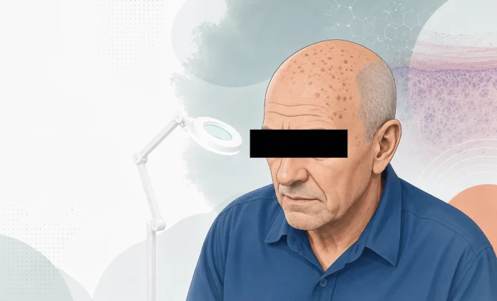
When an actinic keratosis appears, it is almost never alone: around it is a whole field of cancerization, photodamaged skin with invisible lesions. I will explain what it is, why it matters for the risk of squamous cell carcinoma, and how field treatments compare based on two objectives: clearing lesions and preventing cancer.
Basal Cell Carcinoma: The 6 Types and Their Treatment
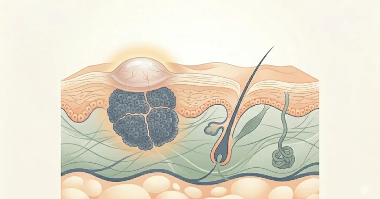
Basal cell carcinoma is the most common skin cancer and also has the best prognosis: it is almost always cured with simple surgery. I will explain its 6 variations, without causing alarm, how it is diagnosed with dermatoscopy, and what treatment corresponds to each.