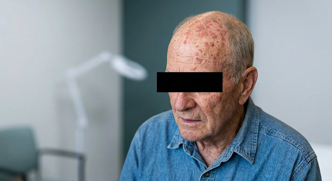
When an actinic keratosis appears on the face, it is almost never alone. Around that rough scab you see is an area of sun-damaged skin with many more altered cells that are not yet visible to the naked eye. That entire territory is called the field of cancerization, and understanding it completely changes how it is treated.
We are discussing a key concept in dermatology related to skin cancer: most squamous cell carcinomas on the face arise precisely where one of these lesions previously existed. In a classic follow-up cohort, approximately 65% of squamous cell carcinomas developed on lesions previously diagnosed as actinic keratoses. (Criscione et al., 2009)
My name is Sebastian Podlipnik and I am Dermatologist at the Melanoma and Skin Cancer Unit of Hospital Clínic de Barcelona. An important part of my work is treating precisely this type of photodamaged skin, and one of the questions I am asked most often in consultation is why I insist on treating "the entire area" and not just the spot the patient has noticed. In this article, I explain why.
The field of cancerization is an area of skin heavily exposed to the sun where, in addition to visible actinic keratoses, there are still invisible damaged lesions and cells. That is why dermatologists talk about treating the entire field and not just the isolated lesion: the risk does not lie in a specific spot, but in the entire terrain.
What is the field of cancerization?
Field cancerization is an area of chronically sun-damaged skin in which visible lesions, such as actinic keratoses, coexist with subclinical cellular damage surrounding those lesions that is not yet visible to the naked eye. It is not an isolated lesion, but an entire territory with a tendency to generate new lesions.
The concept is more than 70 years old. It was first described by a surgeon studying mouth tumors who observed that the healthy-looking tissue around a cancer was already altered at the cellular level, which explained the appearance of multiple independent tumors in the same region. (Slaughter et al, 1953) Decades later, that same idea translated to the skin: areas with cumulative sun exposure develop fields of altered cells that manifest as multiple actinic keratoses, carcinomas in situ, and, in some cases, invasive squamous cell carcinomas. (Willenbrink et al., 2020)
The central idea, and the one I am most interested in having you understand, is this: the problem is not a stain, it is the entire plot of land. That is why it is important to clearly distinguish between the visible lesion and the field that supports it.
- Actinic keratosis
- The visible lesion: a rough, reddish, or scaly patch that can be felt to the touch. It is the "tip of the iceberg" of sun damage.
- Cancerization field
- The entire surrounding area: photodamaged skin with invisible altered cells, from which new lesions may sprout over time.

If you want to delve deeper into the visible lesion itself, I wrote a specific article about What are actinic keratoses. Here we focus on the concept that encompasses them all.
Do you notice several rough lesions in the same area?
I can evaluate the entire area during the consultation, using digital dermoscopy, not just the spot you see.
Request a consultationWhy does it form? The sun and the invisible damage
The field of cancerization is formed by ultraviolet radiation accumulated over years. The sun not only damages the cell that ends up becoming a visible spot, but also leaves its mark on entire groups of cells in the area, generating a "field" of damage that precedes the lesions.
At the biological level, ultraviolet radiation induces mutations in genes that control cellular repair, especially the gene p53 (a "guardian" gene that normally eliminates damaged cells). When that control fails, defective cells accumulate instead of being eliminated and form groups that expand across large areas of sun-exposed skin. That invisible substrate is the origin of both actinic keratoses and carcinomas that may arise afterward. (Wang et al., 2024)

This explains a very frequent experience in the clinic: the patient who is treated for a lesion and, months later, gets two or three more next to it. It is not bad luck or a failed treatment. It is the field, which is still there.
Where does the field of facial cancerization appear?
The field of cancerization appears in areas that have received the most sun over the course of a lifetime. On the face it is very typical, but it is not limited to it: the scalp (especially in people with baldness), the ears, the back of the hands, and the lips are equally common locations.
These areas share a characteristic: they are exposed to the sun almost daily, for decades, often without protection. That is why actinic keratosis is the most frequent tumor lesion in fair-skinned people and usually appears precisely within a field damaged by ultraviolet radiation. (Kandolf et al., 2024)
When the lesion affects the lips, especially the lower one, we speak of a variant with its own name: the actinic cheilitis. And, occasionally, a very thickened and hard lesion known as grows over the field cutaneous horn, which is always advisable to evaluate because there may be a more significant underlying injury.
Is actinic keratosis cancer? The risk of progression
Actinic keratosis is not cancer, but a precancerous lesion. The risk of any single lesion turning into squamous cell carcinoma is low if you look at it in isolation, but it ceases to be negligible when you think about the entire field and the passage of time.
The data help put things into perspective. In a follow-up study of nearly 7,800 actinic keratoses, the risk of a lesion progressing to squamous cell carcinoma was 0.60% per year and 2.57% over four years. The number per lesion is small, but the same study showed the other side of the coin: about 65% of the squamous cell carcinomas in the cohort arose from lesions that had previously been diagnosed as actinic keratoses. (Criscione et al., 2009)

There is a key nuance that I always repeat: you cannot predict which specific lesion will progress or when. Precisely because that crystal ball does not exist, the guidelines recommend treating and monitoring the field rather than waiting to see what happens with each spot. (Kandolf et al., 2024) If you want to better understand how the different types of skin cancer that can arise from this sun damage, I will develop in a separate article.
Are you worried that any of your actinic keratoses might progress? We will evaluate it together.
Request a consultation →How is the field diagnosed?
The diagnosis of the field of cancerization is fundamentally clinical: the dermatologist examines the entire area, not just the lesion that prompts the consultation, looking for that combination of roughness, redness, scaling, and multiple lesions that reveals accumulated sun damage. The key is to look at the terrain, not the spot.
To make a diagnosis, we have imaging techniques that allow us to "see" what the eye cannot capture. Dermoscopy (a magnifying lens with special light) helps distinguish an actinic keratosis from other lesions and detect suspicious signs. In selected cases, more advanced techniques are used, such as confocal microscopy or optical coherence tomography, which explore the skin in depth without the need for a biopsy. When a lesion is suspicious or does not respond to treatment, taking a biopsy is indicated to confirm the diagnosis. (Kandolf et al., 2024)

As a dermatologist dedicated to skin cancer, this overall look is the part I consider most important of the visit: a spot treated without assessing the surrounding field is a missed opportunity. If you notice several rough lesions in the same area, or one that is changing, an in-person evaluation allows exploring the entire area with dermoscopy.
Why treat the entire area and not just the lesion?
It involves the entire field, and not just the visible lesion, because around each spot there is subclinical damage that can also evolve. Treating only what is seen is like cutting weeds while leaving the root: the area continues to produce new lesions. That is why field-directed therapies are generally the preferred strategy.
This is not a personal opinion; it is the framework established by specialized reviews. Field-directed therapies are preferred because they simultaneously address the visible disease and subclinical atypia, whereas single-lesion treatments are reserved for specific cases, such as difficult lesions or when a biopsy is needed. In large fields, the standard approach is to combine both strategies. (Cornejo et al., 2020)
It is worth distinguishing two major strategies, because understanding them will help you read the treatment table that follows.
- Lesion-directed treatment: acts on a specific spot, one by one. The typical example is cryotherapy (freezing the lesion with liquid nitrogen).
- Field-directed treatment: act on the entire damaged area at once, also reaching the invisible damage. This is where creams and photodynamic therapy come in.
Treatments for the field of cancerization: a comparison
The main field-directed treatments are creams (5-fluorouracil, imiquimod, tirbanibulin, diclofenac), topical combinations, photodynamic therapy, and in-office options such as lasers or peels. There is no single "best" option for everyone: the choice depends on the size of the field, the location, each person's characteristics, and, above all, the desired goal.
And here is the idea I want you to take away, because it changes how any comparison is read: a field treatment is judged by two distinct objectives that are not interchangeable.
- Goal 1: clear the lesions (clearance)
- Make visible actinic keratoses disappear. That is what most studies measure.
- Objective 2: reduce squamous cell carcinoma
- To make less cancer appear over time. That is the underlying goal, and only a few treatments have demonstrated it.
When it comes to the ability to clean wounds, almost all options work similarly. The benchmark comparative trial directly compared four first-line treatments for multiple keratoses of the head: At 12 months, 5-fluorouracil was the most effective, with 74.7% of patients showing no treatment failure, compared with 53.9% for imiquimod and 37.7% for photodynamic therapy. (Jansen et al., 2019) Reviews comparing all modalities find them quite similar in clearance, with diclofenac being the weakest. (Worley et al, 2022)
In reducing squamous cell carcinoma, on the other hand, the list becomes much shorter. Topical 5-fluorouracil was the first to demonstrate this: in the VAKCC trial, published in JAMA Dermatology, a single course of treatment on the face and ears reduced the risk of squamous cell carcinoma by 75% during the first year (1% versus 4% in the control group), although the effect was transient and specific to squamous cell carcinoma. (Weinstock et al., 2018)

A cohort of about 5,700 patients subsequently supported this: 5-fluorouracil was associated with fewer keratinocytic carcinomas than imiquimod. (Neugebauer et al, 2019)
The most promising results come from combining 5-fluorouracil with calcipotriol, a vitamin D derivative. In a randomized trial, this combination, applied for only four days, reduced actinic keratoses by 87.8%, compared with 26.3% for 5-fluorouracil alone. (Cunningham et al., 2017) And, most importantly in terms of what really matters, at the three-year follow-up, patients treated on the face and scalp developed significantly fewer squamous cell carcinomas (7% versus 28%), an effect that appears to be linked to a lasting immune response from the skin itself. (Rosenberg et al, 2019) It is a promising result, still in studies of modest size, but it fits with the idea of not only clearing the field, but teaching the immune system to monitor it.
The following table summarizes all the options according to those two objectives.
| Treatment | How it acts | Objective 1: clean lesions | Objective 2: Does it reduce squamous cell carcinoma? | Expected reaction | Oriented to |
|---|---|---|---|---|---|
| 5-fluorouracil (cream) | Topical chemotherapy that destroys damaged cells throughout the area. | High dose: the most effective over the course of a year in the comparative trial (74.71 TP3T without failure). | Yes, transient: 75% fewer basal cell carcinomas in the first year. | Inflammation and crusts for several weeks. | Fields with many injuries; when prioritizing prevention. |
| 5-fluorouracil + calcipotriene (cream) | Add calcipotriol (vitamin D) to 5-fluorouracil to activate local immunity. | Very high with a short duration: 87.81 TP3T reduction in injuries over 4 days. | Yes, the most lasting sign: fewer squamous cell carcinomas at 3 years (7% vs. 28%). | Marked redness, but without the crusts or ulcers of classic 5-fluorouracil. | Facial field and scalp; when the goal is to prevent the squamous cell carcinoma. |
| Imiquimod (cream) | Immunomodulator: activates local defenses against altered cells. | Annual average (53.91 TP3T without failure). | Clearance data only; not demonstrated. | Redness and scabs; sometimes mild flu-like discomfort. | Mild fields; alternative to 5-fluorouracil. |
| Tirbanibulin (ointment) | Halts the division of atypical cells; short regimen in studies. | Complete remission of 44-54% after 2 months; recurrence of 47% after one year. | Clearance data only; not demonstrated. | Mild and transient redness and peeling. | Small fields; when comfort is prioritized. |
| Diclofenac (gel) | Long-term topical anti-inflammatory. | The lowest of the field options (around 23%). | Clearance data only; not demonstrated. | Good tolerance, but long treatment (2 to 3 months). | Mild cases or those who cannot tolerate more intensive treatments. |
| Photodynamic therapy (PDT) | A product sensitizes damaged skin and then illuminates to destroy it. | Good initial response; lower durability after one year (37.7%). | Signal only with repeated sessions at high risk; no definitive proof. | Redness and stinging during the session; scabs for a few days. | Extensive areas on the face and scalp; in-office treatment. |
| Laser or spot peeling | They renew the surface layer of the entire area (fractional ablative laser or chemical peel). | Comparable to creams and photodynamic therapy. | Clearance data only; not demonstrated. | Redness, peeling, and crusting depending on depth. | Heavily photodamaged skin; can be combined with other treatments. |
Regarding tirbanibulin, the most recent drug, it is important to have realistic expectations. In its Phase 3 trials, it achieved complete clearance of lesions in approximately 44% and 54% of patients after two months, with the advantage of a short treatment regimen. However, nearly half of those who achieved clearance (47%) experienced a recurrence of lesions within a year. (Blauvelt et al, 2021) It is a reminder that no treatment "cures" the field permanently.
You will see that there are no prices or specific guidelines in the table, and that is deliberate. What treatment is appropriate, for how long, and with what frequency is a decision that depends on each case and is up to the dermatologist's evaluation. My goal here is for you to understand the options, not for you to self-treat. If you have an area that you think needs treatment, I can guide you in a online visit or explore it in face-to-face consultation.
Prevention and monitoring: the field does not disappear
Treating the field is not the end of the road, but the beginning of follow-up. The field of cancerization persists even if the lesions clear, which is why photoprotection, self-examination, and periodic check-ups are as important as the treatment itself. The damaged skin is still there, and it should be cared for in the long term.
Guidelines insist that prevention is part of treatment. In fact, in the US clinical practice guideline on actinic keratosis, protection against ultraviolet radiation appears as a strong recommendation, at the same level as the most effective treatments. (Eisen et al, 2021) In high-risk individuals, European recommendations add self-examination and repeating field treatment in previously affected areas. (Kandolf et al., 2024) This is of particular importance in immunosuppressed or transplant patients, in whom actinic keratoses progress more easily and early field treatment is a priority. (Heppt et al., 2019)
There is also a complementary approach supported by strong evidence, particularly for those who have already had skin cancer: chemoprevention with nicotinamide. In a randomized trial conducted among people who had had at least two cases of non-melanoma skin cancer, oral nicotinamide (vitamin B3) reduced the incidence of new carcinomas by 23% compared to placebo after one year, with a reduction of nearly 30% observed for squamous cell carcinoma. (Chen et al, 2015) It is precisely the patient profile with an active field cancerization and a history of lesions, and I develop it in detail in my article on the nicotinamide as chemoprevention for skin cancer. An honest nuance is in order: the benefit is maintained while taking it, and in transplant patients a subsequent trial did not find that same protection. (Allen et al, 2023)
"We treat the field of cancerization to prevent skin cancer, not for aesthetics. That is why the visit does not end when a spot disappears: follow-up is what truly protects your skin in the long term."
Dr. Sebastian Podlipnik
Frequently asked questions about the field of cancerization
What is field cancerization in simple terms?
Is actinic keratosis cancer?
Why do we need to treat the whole area and not just the visible spot?
What is the best treatment for field cancerization?
Does the field of cancerization heal completely?
Do you notice several rough lesions or a changing spot in a sun-exposed area? I can help you assess the entire field, not just the lesion.
Online Visit On-site VisitReferences
- Slaughter DP, Southwick HW, Smejkal W. Field cancerization in oral stratified squamous epithelium. Cancer, 1953. PubMed
- Willenbrink TJ et al. Field cancerization: Definition, epidemiology, risk factors, and outcomes. Journal of the American Academy of Dermatology, 2020. JAAD
- Cornejo CM et al. Field cancerization: Treatment. Journal of the American Academy of Dermatology, 2020. JAAD
- Criscione VD et al. Actinic keratoses: Natural history and risk of malignant transformation in the VATTC Trial. Cancer, 2009. PubMed
- Jansen MHE et al. Randomized Trial of Four Treatment Approaches for Actinic Keratosis. New England Journal of Medicine, 2019. New England Journal of Medicine
- Blauvelt A et al. Phase 3 Trials of Tirbanibulin Ointment for Actinic Keratosis. New England Journal of Medicine, 2021. New England Journal of Medicine
- Weinstock MA et al. Chemoprevention of Basal and Squamous Cell Carcinoma With a Single Course of Fluorouracil, 5%, Cream (VAKCC Trial). JAMA Dermatology, 2018. JAMA Dermatology
- Neugebauer R et al. Comparative effectiveness of treatment of actinic keratosis with topical fluorouracil and imiquimod in the prevention of keratinocyte carcinoma. Journal of the American Academy of Dermatology, 2019. PubMed
- Cunningham TJ et al. Randomized trial of calcipotriol combined with 5-fluorouracil for skin cancer precursor immunotherapy. J Clin Invest, 2017. PubMed
- Rosenberg AR et al. Skin cancer precursor immunotherapy for squamous cell carcinoma prevention. JCI Insight, 2019. PubMed
- Worley B et al. Treatment of actinic keratosis: a systematic review. Archives of Dermatological Research, 2022. PubMed
- Liew YCC et al. Photodynamic therapy for the prevention and treatment of actinic keratosis/squamous cell carcinoma in solid organ transplant recipients: a systematic review and meta-analysis. J Eur Acad Dermatol Venereol, 2020. JEADV (Wiley)
- Chen AC et al. A Phase 3 Randomized Trial of Nicotinamide for Skin-Cancer Chemoprevention (ONTRAC). New England Journal of Medicine, 2015. New England Journal of Medicine
- Allen NC et al. Nicotinamide for Skin-Cancer Chemoprevention in Transplant Recipients (ONTRANS). New England Journal of Medicine, 2023. New England Journal of Medicine
- Eisen DB et al. Guidelines of care for the management of actinic keratosis. Journal of the American Academy of Dermatology, 2021. PubMed
- Kandolf L et al. European consensus-based interdisciplinary guideline for actinic keratosis, Bowen's disease and field cancerization. J Eur Acad Dermatol Venereol, 2024. JEADV (Wiley)
- Wang Z et al. Actinic keratosis pathogenesis: current state and perspectives. Front Med, 2024. Frontiers in Medicine
- Heppt MV et al. Local interventions for actinic keratosis in organ transplant recipients: a systematic review. Br J Dermatol, 2019. British Journal of Dermatology
- European Medicines Agency (EMA). EMA suspends Picato (ingenol mebutate) as a precaution while review of skin cancer risk continues. Press release, January 17, 2020. EMA