
Skin cancer is the most common type of cancer and can affect up to 1 in 5 people by the age of 70. The most worrying thing is that we continue to see a progressive increase in cases over the last decades that persists despite all the prevention campaigns that we dermatologists carry out at the population level. (See incidence study in catalonia) So why and how to detect skin cancer early?

Unlike other neoplasms that develop inside the body, skin cancer is formed in the outermost layer of the skin and is often visible. That is why regular skin examinations, both at home and with a dermatologist, are especially important. The good news is that most cases of skin cancer are curable if diagnosed and treated early.
Self-examination by the patient
La early detection of skin cancer saves lives. And that's why learning to examine your own skin gives you the power to detect suspicious lesions at very early stages, before skin cancer becomes dangerous, disfiguring or deadly. In several scientific studies we have seen that women self-detect more than half of melanomas. In addition, melanomas detected by women have a better prognosis than those detected by men because they were identified at an earlier stage. Therefore, we see how early detection by the patient can drastically change the prognosis.
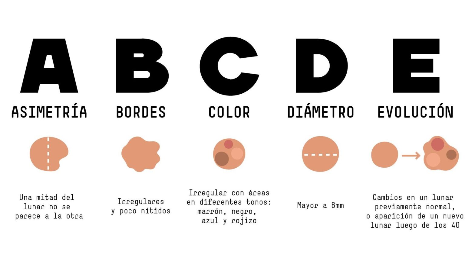
How to detect skin cancer can be easy with a few simple tips. The points of the ABCDE is a mnemonic rule that will help patients to identify suspicious lesions more easily while checking their moles. Thus, when a mole with the following characteristics is seen, it should be considered suspicious and a specialist should be consulted:
- Asymmetry of a mole on any of its axes
- Birregular orders
- Codor varied, not uniform
- DDiameter greater than 6 mm
- Evolution. If we detect that one of our moles is changing during the time
What do dermatologists do in the office for skin cancer screening?
For many years dermatologists have been teaching our patients how to detect skin cancer early using the ABCDE rule. However, we have much more precise tools that allow us to detect changes before the melanoma shows ABCDE features. Below I will show you some of the techniques we use during the consultation to improve our diagnostic yield.
Dermatoscopy
La dermatoscopy or epiluminescence refers to the direct examination of the skin by means of a high magnification lens and polarized light that allows us to see structures through the skin. It is an essential tool in the consultation of all dermatologists that allows us to differentiate early skin cancer. Dermoscopy is mainly used to evaluate pigmented lesions such as moles and melanoma but it also has many other uses.
Thanks to dermoscopy we have the possibility of detecting skin cancer in very early stages. Here are some images of melanoma where you can see in detail what this type of cancer looks like.


Digital nevus tracking
New diagnostic computer systems make it possible to obtain total body maps, locate nevi and electronically archive images of lesions with digital epiluminescence microscopy. This makes it possible to perform very precise serial controls of the patient and to detect minimal changes suggestive of malignancy even when there are a large number of lesions. Watch the following video and see how we dermatologists perform the digital follow-up of moles.
Confocal microscopy
Sometimes there are situations in which it is very difficult to differentiate an atypical (dysplastic) mole or an incipient melanoma. Or also, it often happens that there are lesions in areas that are difficult to explore or where a biopsy can leave significant scarring (such as on the face). In these cases, we can using confocal microscopy which allows us to obtain cellular resolution images in a non-invasive way. Thanks to confocal microscopy we can be even more precise in the diagnosis of our patients and thus avoid unnecessary surgeries.

Do I need to have a mole check?
It is recommended that if you have never had a mole check-up, you should have it done by an expert dermatologist to see the characteristics of your nevi. Depending on the individual risk factors found and your family history, your dermatologist will make an individualized recommendation for follow-up. In the following link I leave you more details of how we perform the first visit of moles or nevi in our practice.
What can I do to prevent skin cancer?
The good news is that skin cancer is preventable, and in case it appears and is detected early it can be cured. To protect yourself, here are some tips:
- Avoid the sun during peak hours: UV radiation tends to be much higher between 12:00 and 16:00 (depending on latitude), so avoid exposure to the sun during these hours.
- Use sunscreen all year round: Although during the autumn and winter months the sun is not as strong, we continue to receive UV radiation that penetrates the clouds. Skin cancer is not only caused by intense summer exposure, but UV radiation accumulates throughout our lives, progressively increasing the risk of skin cancer. Check out the best sunscreen creams.
- Avoid tanning beds: tanning beds or booths emit a high dose of UV radiation. There are now numerous studies showing a higher incidence of melanoma in young patients who have been tanning booth users during adolescence.
- Wear clothing with UV protection: For some years now, many international brands have been introducing UV sunscreens in their fabrics.
- Wear sunglasses: look especially for those that block UVA and UVB rays.
- Check your skin regularly and report changes to your dermatologist as soon as possible.
- Vitamin D is not an excuse: Many of my patients argue that they need vitamin D and therefore expose themselves for hours in the sun. But more sun exposure does not mean more vitamin D.
What does the future hold for early detection of skin cancer?
Surely in the future we will have automated equipment in 3 dimensions and with artificial intelligence that will allow us to automate many of the functions we do today. Below is a video of how some of these devices could be used in our clinical practice.


8 Responses
Good afternoon, about three months ago a small mole appeared between my index finger and thumb, today I saw it again and it is a little bigger, in addition, another mole of the same size appeared next to it. The truth is that I don't have many moles and it seems a little strange to me, but I don't know if I should worry and go to a specialist.
Hi Sharon,
Undoubtedly from what you say I think it would be important to be evaluated by a specialist. If you have a new mole during adulthood and it is growing, it is preferable to have it checked by a specialist.
Best regards
Good afternoon
A question: How can I remove warts on my neck at home?
Dear Arturo,
I never recommend removing skin lesions without first having them evaluated by a specialist. Here is an explanation in an article on why.
The information provided here is very reliable and truthful. Very good blog.
Dr. Roberto Morrison.
Dermatologist.
Thank you very much Roberto for your comment.
I'm glad you like the content of the Blog and I hope to continue posting things that may be of interest to you.
Greetings
Very good morning, I have a mole on my forehead and I would like to remove it, how much would it cost me and where is your office?
Hello Jhonny, my The practice is located in Barcelona, Spain. To know the price it is necessary to first assess your mole and see what type it is, what size it is and the complexity of removing it. Depending on these things usually a budget is made. Best regards