Malignant moles or melanoma How to identify and treat them?

By Dr. Sebastian Podlipnik

Moles or nevi are common and present in most people. They can appear anywhere on the body and are, for the most part, harmless. While it is normal to have moles there are also malignant moles, cancerous moles or melanoma.  

Fortunately, there are different signs and symptoms that help us to understand the can help to identify them and promptly consult a dermatologist if necessary. 

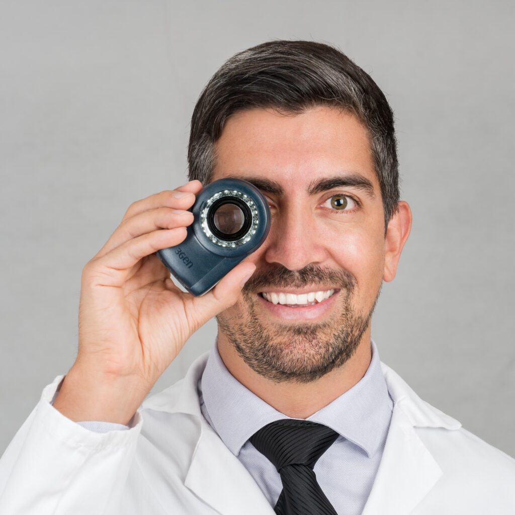
Dr. Sebastian Podlipnik holding a dermatoscope

My name is Sebastian Podlipnik and I am Dermatologist at the Melanoma and Skin Cancer Unit of Hospital Clínic de Barcelonaone of the world's most renowned centers for the treatment of skin cancer. 

Annually I see many patients with cancerous moles of the melanoma type and I believe that many times they could have been diagnosed earlier. In this article I will give you some clues so that you can make an early diagnosis.

What will this article cover?

Melanomas or malignant moles, what are they?

When we talk about malignant moles or cancerous moles we are referring to malignant melanoma. The melanoma is a malignant tumor that develops from melanocytes. whose main function is the production of melanin that gives color to our skin. Although malignant moles almost always appear on the skin, they can also appear on the eye, lips, oral mucosa or even genital mucosa.

Although melanoma accounts for only 5% of all melanomas, it is not a different types of skin cancer that can affect us, produces more than 90% of the deaths attributed to skin cancer. Ref This is why its early identification is so important.

In the illustration we can see how the following processes necessary for the development of a malignant mole occur. 

  1. The melanocytes that are chronically exposed to UV radiation are acquiring mutations
  2. Eventually, some of these mutations can trigger melanoma
  3. Now the malignant mole cells can grow superficially uncontrollably. At this point we may be able to detect melanoma early.
  4. However, if we do not consult these cells can invade deeply into the skin in a process that can last for months.
  5. Finally, if the cells reach the lymphatic or bloodstream, the following can occur metastases in other organs

Do all melanomas arise on moles?

From infancy to about age 40, our skin produces between 10 and 50 moles, which are basically clusters of pigmented cells or melanocytes. In general, these moles are harmless. But what percentage of melanomas come from moles?

During the last few years we have seen in different scientific studies that only 26% of melanomas appear on the moles.In fact, it is rare for melanoma to arise from a pre-existing mole.Ref A recent theoretical study has shown that for a 20-year-old individual, the lifetime risk of any individual mole developing into melanoma by the age of 80 years is approximately 0.03% (1 in 3,164) for men and 0.009% (1 in 10,800) for women.Ref

iconography showing the percentage of melanomas or malignant moles that develop on top of moles

This is very important because many of my patients tell me to remove their moles to prevent the risk of developing melanoma. However, preventive mole removal does not really reduce the risk at all. of developing a malignant mole. 

The problem is that when a melanoma begins to develop it can look very much like an ordinary mole. If you have a lot of moles and some of them have atypical characteristics, what you should do is to have a specific monitoring of moles with your dermatologist.

How serious is it to have melanoma?

It depends! Melanoma is a malignant mole that if detected in advanced stages can be fatal. However, there is a 99% survival rate when the disease is detected in its early stages.Ref And one of the best ways to make sure that happens is with a periodic self-examination of our skin or in case you have many moles doing and regular control with your dermatologist. Ref

Early detection makes all the difference

99%

Survival at 5 years of age in patients who are found to have melanoma in early stages. This percentage drops to 66% if the disease reaches the lymph nodes and a 27% if there are metastases in other organs

What are the symptoms of malignant moles?

In the early stages melanomas may not give any symptoms and in fact may look very much like benign moles. That is why one of the most important signs is the change your moles may have. Dermatologists recommend checking tCheck your skin every month for new moles or irregularities. If you detect a change that persists over time, it may be a sign of incipient skin cancer.

I recommend that you examine your skin from head to toe every month. It is a simple but very effective way to detect a malignant mole early.

Now I will leave you with other tips and recommendations so that you can identify a melanoma early and you can visit your dermatologist if necessary. 

Sign of the ugly duckling

The ugly duckling sign is a very useful warning sign of melanoma. This recognition strategy is based on the concept that most normal moles on the body look similar to each other, while the melanomas stand out like ugly ducklings in comparison (different)..Ref 

This highlights the importance of not only checking for irregular moles, but also comparing any suspicious spots with surrounding moles to determine if they look different from their neighbors.

Back of a woman with malignant moles with the sign of the ugly duckling

These ugly duckling lesions or atypical lesions may be larger, smaller, lighter or darker compared to surrounding moles. Isolated lesions with no surrounding mole for comparison are also considered ugly ducklings.

ABCDE of melanoma

The ABCDE rule is another mnemonic rule that allows us to identify changes in freckles and moles that may be suggestive of melanoma.Ref 

  • A is for Asymmetry: One half of a mole is different from the other half
  • B for Border: The spot or mole has irregular edges
  • C for Color: The lesion has different shades of brown or black, or sometimes with pink, red, white or blue spots.
  • D is for Diameter: The mole is more than 6 millimeters in diameter, although sometimes melanomas can be smaller.
  • E for Evolution: The mole is changing in size, shape or color.

It is important to report any signs of ABCDE to your dermatologist. If you see, for example, a change or new spots on your skin, or moles that look different from the rest, it is important to consult your dermatologist. 

Other signs of suspicion of malignant moles

Identifying a possible cancerous mole is not easy, and not all melanomas follow the rules I have shown you previously. Some of these cases are:

  • Amelanotic melanomas lack the dark pigment or melanin that gives color to most moles. Amelanotic melanomas can be pinkish, reddish, white, skin-colored or even clear and colorless, making them difficult to recognize. Fortunately, they are rare, but if you have a red lesion on your skin that bleeds, increases in size or is bothersome, it is important to consult.
  • Acral lentiginous melanomaThe most common form of melanoma in people of color. It usually appears in places that are difficult to detect, such as under the fingernails or toenails, on the palms of the hands or on the soles of the feet.

Finally, I will leave you with a few last clues that can help you identify a malignant mole in time, such as when:

  • A wound that will not heal
  • There is a spread of brown or black pigment from the edge of a blemish into the surrounding skin.
  • There are symptoms such as itching, tenderness or pain over a mole.
  • Changes appear on the surface of a mole such as scaling, oozing, bleeding or the appearance of a lump or bump.

When to see a dermatologist?

In short, we must visit a dermatologist when any new moles or freckles appear on your skin. In addition, it is important to consult if a wound that does not heal despite adequate treatmentor when any pre-existing mole begins to change (growing, swelling, itching). Finally, it is very important to be aware of any spots, moles or lesions on the skin. unusual feature (ABCDE rule or ugly duckling)

Be sure to show your dermatologist at consultations any areas that concern you and ask him or her to look at any areas that are difficult for you to see. Sometimes it is difficult to distinguish between a melanoma and a normal mole, even for dermatologists, so it is important to show the dermatologist any moles you are unsure about. 

Request an appointment

Have you been diagnosed with skin cancer and have doubts?

Photos of malignant moles

Melanomas can take many different forms and will not always follow the rules. Now that you know the signs to be concerned about, I will leave you with some images of malignant moles to reinforce the ideas previously presented. 

Malignant mole or melanoma on a patient's chest

Invasive melanoma

Young male presenting with an ugly duckling sign lesion and progressive growth

Malignant mole or melanoma on a patient's shoulder

Incipient melanoma

Young man with progressively growing pigmented lesion on the back with the ugly duckling sign

Melanoma or cancerous mole on the forearm of a female patient

Incipient melanoma

Fast-growing pigmented lesion on the left forearm that is larger than all the rest.

Invasive melanoma on a patient's thumbnail

Melanoma on the nail

Brown pigment can be seen around the nail and destruction of the nail plate.

Amelanotic melanoma or malignant unpigmented moles growing on a patient's back

Melanoma amelanotic

In this case I show you a melanoma without pigment that grows as a red "wart".

acral melanoma or malignant mole on the sole of the skin which looks like a black spot

Acral melanoma

Black spot on the sole of the foot that grew progressively over the years until the patient became aware of it. It is a place of difficult visualization.

Note: Not all melanomas look the same! These photos serve as a general reference of what a melanoma may look like. If you see something NEW, CHANGING or UNUSUAL on your skin, get checked by a dermatologist.

What are the risk factors for melanoma?

There are different risk factors that can increase the chances of developing a cancerous mole. Many of these factors are genetic, but the most important ones that we can prevent are environmental.Ref

Genetic and phenotypic factors

  • Genetic risk factors (genotyping) are those that are found within our genes and that we cannot control. When in a family there are several members with melanoma, or the same person has had more than 2 melanomas, we study their genes to find out if there is a predisposition to develop melanoma.
  • The phenotype refers to the characteristic features of each person such as skin color and the number of moles we have. The combination of these factors allows us to identify at-risk populations that are more likely to develop melanoma.

There is not much we can do about these factors to modify the risk of developing melanoma. But we can select patients who will require a specific follow-up with the dermatologist.

Infographic showing genetic risk factors for melanoma or malignant moles.
If the patient has more than one of these factors the risk is multiplied.

Environmental factors

On the other hand, we have the environmental factors (exposome) which refers to all the external causes that influence our skin and can favor the development of a cancerous mole. Among the most important causes we have:

  • Intense intermittent exposure to UV radiation. For example, when we become red after intense sun exposure during the vacations and the skin subsequently flakes off.Ref
  • Chronic exposure to UV radiation. For example, in people who are constantly exposed to the sun.Ref
  • Use of tanning booths, especially in people under 35 years of age, has been found to increase the risk of melanoma by 2 times. In addition, a recent study has shown that just 10 sessions of UVA can increase the risk of developing melanoma by 1.7 times.Ref
  • Immunosuppression. In patients who for some reason are immunosuppressed. We see it a lot in organ transplant patients who have to take immunosuppressants.Ref
 

Download my free photoprotection guide

Image of the cover of the sun protection guide

Access my complete guide to photoprotection, where I share all my exclusive tips and recommendations - get it now!

What to do to prevent the appearance of malignant moles?

There is not much we can do to modify genetic factors. However, in the face of environmental factors, we can do a great deal to prevent the appearance of a malignant mole and also maintain a healthy and youthful skin.Ref 

Of all the measures we can do, sun protection and avoiding tanning booths are the 2 most important. If you want to become a expert in photoprotection I leave you this article to see the best ways to protect yourself. 

On the other hand, identifying patients at risk allows us to improve the efficacy and efficiency of follow-up programs and thus detect very early melanomas. In addition, at the individual level, it allows us to improve clinical decisions including periodic follow-ups, digital follow-ups, or the performance of biopsies of suspicious lesions. 

Do all melanomas have the same prognosis?

When detected and treated early, melanomas are highly curable and many times simply removing the suspicious mole will suffice. However, as time goes on the melanoma will continue to develop until it becomes invasive.

In order to know the prognosis of patients with melanoma, we use the classification of the American Joint Committee of Cancer (AJCC)  that classifies patients broadly speaking from stage 0 to stage IV.Ref

Infographic showing the stages of melanoma or malignant moles.
image obtained from sciencedirect.com

Once we diagnose a melanoma and do all the initial studies we will be able to perform staging. The stage provides the physician and the patient with a common language to understand how advanced the cancer iswhere it is located and what treatment options are available. I will now explain in general terms what the different stages of melanoma mean: 

  • Stadium 0: It is also called melanoma in situ. It means that there are melanocyte cells located only in the epidermis, that is, the most superficial layer of the skin. This is the earliest stage. It is best to detect and treat melanocytes here before they have a chance to become invasive.
  • Stage I: In general, a stage I melanoma has grown slightly larger in the skin but is still quite superficial. Survival is excellent in these cases. 
  • Stage II: A stage II melanoma has grown deeper into the skin and has more high-risk features, but has not yet spread to the lymph nodes or the rest of the body. 
  • Stage III: A stage III melanoma diagnosis means that the cancer cells have spread to nearby lymph nodes, but not to distant organs or any other part of the body.
  • Stage IV: In stage IV, melanoma has spread to other organs and/or distant lymph nodes.

Treatment and follow-up of malignant moles

Treatment and follow-up of melanoma has to be individualized in each case depending on the stage of the melanoma. In early cases only surgery will be necessary to remove the superficial melanoma cells. However, in advanced stages, in addition to surgery, it may be necessary to use immunotherapy or target molecules if the neoplasm has spread beyond the skin. 

If you are interested in knowing more about melanoma treatment I can write an article about it. Leave me a comment and I will know if it might be of interest. 

Final tips for detecting malignant moles

Regardless of your risk of developing melanoma, examine your skin from head to toe once a month. Look for existing moles or lesions that grow or change.

Keep in mind that if you have many risk factors and atypical moles, monthly self-exams may not be sufficient. See your dermatologist at least once a year for a professional skin examination.

If you have had a melanoma, you need to follow up regularly with your dermatologist. 

Share this article

43 Responses

  1. Hello doctor, I am 25 years old and I am quite light skinned and I have quite a few moles. Recently (2 weeks ago) I feel a kind of "tingling" in the area of one of my moles on my back. It is intermittent but I do notice it from time to time. It is nothing more than tingling and, although the mole is large, I have had it for some time and it does not show any signs of ABCD testing. Can this tingling be a cause for concern?

    1. Hello Álvaro,

      Tingling in a mole can have several causes, and is usually not a cause for concern. However, I would recommend that you consult a dermatologist or physician for an in-person evaluation. They will be able to determine the exact cause and provide you with more information. Best regards

  2. Hi! My dad, who is 57 years old today, has had a mole on his face since birth, several in the family have had it. It is lumpy and smooth and has a few long hairs growing on top of it. Over the years it has been growing like the others in the family, today it is the size of a pencil eraser. Yesterday I notice that it was inflamed, and today he says that it itches and hurts the skin around it, the mole has not changed color, nor does it bleed, nor is it cracked, nor is it irregular. We are quite concerned, but unfortunately he is in a fairly remote part of town to go to the doctor today, he plans to go next week. I am very concerned, what do you think it is. I understand you can't make a diagnosis but I want some information. I have been looking for such a mole on the internet and found nothing. Thank you for your attention.

    1. Dear Cintia, it is probably nothing important but it would be a good idea to consult a dermatologist. When we have such rapid changes of inflammation, it is usually an inflamed nevus. Best regards

  3. Hello Doctor: I am writing to you from Santiago de Chile. In 2009 I had a melanoma in situ and over time the removal of several atypical nevi. Now in my last checkup, having had laser hair removal on my legs, my dermatologist noticed many changes in my moles, so much so that 4 were removed because they looked suspicious. I still do not have the result of the biopsy, but my question is if there are studies regarding the malignization of nevi by the laser or it is about changes to the dermoscopy that could generate a suspicious appearance. I have read some posts on the subject, and have not found any association between laser and malignant changes in moles after laser depilation but for now I have forbidden it. I appreciate your comments.

    1. Hi Fabiola, I hope you are very well. So far we can conclude that it does not seem that the use of laser or intense pulsed light (IPL) for cosmetic hair removal is associated with an increased risk of melanoma. What is true is that if they have lasered your nevi, they will show changes that may appear suspicious. Therefore, the best recommendation is to always protect your moles with a sheet or white paper so that the hair removal laser cannot affect them.

  4. I had always had a big mole on my back, but three years ago when I was pregnant it grew; a dermatologist saw it and said it was not malignant; in May my family doctor gave me cold nitrogen and it did not fall off completely as it was big, he came back and applied it, but it did not fall off; now the skin is a little pink around it and the mole has two black spots, it burns or sometimes itches....

  5. Hi, I have a mole that started to hurt when I felt it, the first day it swelled, but now it doesn't, it only hurts a little when I touch it.

  6. A mole that I have had since I was a child has changed, first it had a black spot in the center, then the spot disappeared, but now it itches a lot, I almost always have itchy discomfort, and I get hairs on the mole, which I did not have before, I have already made an appointment at my medical center, but it scares me a little.

    1. Dear Evelyn, from what you tell me it is probably due to an inflamed mole but you don't have to worry too much. In any case, you should go to a dermatologist to have it checked.

Sebastian Podlipnik - Skin cancer

Sebastian Podlipnik

Dermatology Blog

I am a dermatologist and cum laude PhD and author of multiple research studies. I specialize in skin cancer, laser technologies and longevity in dermatology. The intention of this blog is to bring you closer to topics of interest in dermatology and research.

My favorites
Read my book
Subscribe

And get exclusive and quality information for my readers.

Subscribe to my newsletter

To stay informed about the latest news and I will send you my free photoprotection guide