"Flesh mole" is not a medical diagnosis; it's a catch-all. People use that colloquial name for almost any skin-colored bump, so under that label fits a long list of lesions. In this article, I focus on the five most commonly confused with that term: almost all are benign, but one can disguise skin cancer.
If you have made it this far looking for how to remove a flesh mole, the first thing is to know which of the five you have, because the method changes completely depending on the lesion. Have you ever wondered why some are removed with scissors and others with a laser? Here you will understand.
My name is Sebastian Podlipnik and I am Dermatologist at the Melanoma and Skin Cancer Unit of Hospital Clínic de Barcelona. In my practice, I see patients daily who call very different things "flesh moles," and a good part of my work is to properly identify each bump before deciding what to do with it.
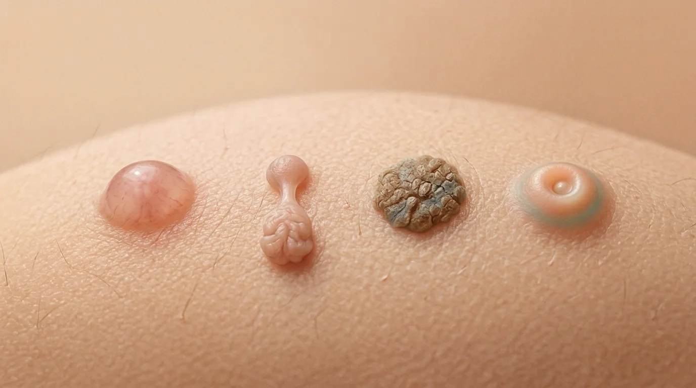
"Flesh-colored mole" is a popular term that people apply to many skin-colored lesions. The five most common are intradermal melanocytic nevus, acrochordon or soft fibroma, clear seborrheic keratosis, sebaceous hyperplasia, and dermatofibroma. Almost all of them are benign, but only a dermatologist can confirm this.
What is a "flesh mole" really?
A "flesh-colored mole" is a common term, not a medical condition. People use it to describe any bump or lesion that is the same color as the skin, without any obvious brown pigmentation, so it actually encompasses many different conditions. In this article, I focus on the five most common ones, and only one of them is a true mole in the medical sense.
In medicine, the term "lunar" is reserved for lesions formed by melanocytes, the cells that give the skin its color. Of the five we'll discuss here, only the intradermal melanocytic nevus (a mole whose cells are located in the deep layer of the skin) fits that definition. The other four originate from different tissues: a band of collagen, an enlarged sebaceous gland, an accumulation of keratin, or a proliferation of fibroblasts.
That's why grouping them under the same name causes so much confusion, which is why the treatment for each one is completely different from that of the others. Distinguishing them at a glance is difficult even for a trained eye: I often need a dermatoscope—a lens with polarized light that magnifies the lesion—to be sure.
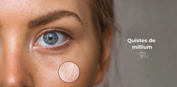
There are other skin-colored lesions that could also fall into this category, such as milia or small epidermal cysts, but they are less common or go by other common names. Here, I’ll focus on the five that I most often hear referred to as "flesh-colored moles" in my practice, ranked from the most "authentic" to the most commonly confused. Keep reading—we’ll start with the only one that’s actually a mole.

Not sure which of the five is right for you?
I can tell you during the consultation, using digital dermoscopy, in a single visit.
Request a consultationIntradermal nevus: the most authentic "flesh mole"
The intradermal melanocytic nevus is the mole that most deserves the name "flesh mole." It is a benign, dome-shaped, and soft lesion, usually skin-colored or slightly brownish, that appears mostly on the face and neck. It tends to grow slowly over years, and many lose their pigment over time.
These moles are also known by two classic names based on their shape: the Unna (flatter and wrinkled, like a blackberry) and the Miescher's nevus (more dome-shaped and firm, typical of the face). In a series of 202 intradermal nevi confirmed by biopsy, the most common vascular pattern observed under the dermatoscope was “comma-shaped vessels”—curved capillaries that help distinguish them from malignant lesions such as basal cell carcinoma or amelanotic melanoma. (Greco et al., 2020)
The vast majority are harmless and do not require treatment. Many people want to have them removed for cosmetic reasons, especially when they rub against clothing or glasses, and that is perfectly reasonable. The important thing is that, before removing a raised mole, a dermatologist confirms that it is indeed benign. If you’ve developed a new one as an adult or notice that it’s changing, you’ll want to read about moles that appear in adulthood and when it is advisable to monitor them.

Acrochordon or soft fibroma: the little skin tag
An acrochordon, also known as a soft fibroma or "fibroepithelial polyp," is that small, skin-like tag that hangs from the neck, armpits, eyelids, or groin. It is skin-colored or slightly darker, soft, and is usually attached by a thin stalk. It is completely benign and very common from middle age onwards.

Its pendulous, smooth appearance distinguishes it from a viral wart, which is rougher, flatter, and sometimes has black dots (small thrombosed capillaries). Warts are caused by the human papillomavirus and can be contagious, whereas skin tags are not contagious at all. If you are in doubt between one thing and another, I clarify it in the article about How to Remove Warts and how it differs from other injuries.
They result from a combination of repeated friction, genetic predisposition, and hormonal and metabolic factors. That’s why they’re so commonly found in skin folds, where the skin rubs against itself or against clothing. And here’s a detail that almost no one knows—and which, to me, is one of the most interesting aspects of this entire group of skin lesions.
Why can skin tags be a metabolic clue?
Having a lot of skin tags can be more than just a cosmetic issue: several studies link them to insulin resistance and metabolic syndrome. They aren't dangerous in and of themselves, but when they appear in large numbers, they may be a sign of abnormalities in blood sugar, lipids, or blood pressure—conditions it's important to be aware of.
In a case-control study, patients with skin tags were much more likely to have diabetes and worse glucose, cholesterol, and triglyceride levels than people without them. (Shah et al., 2014) This finding has been replicated in a European population: in a cohort of adults, skin tags on the neck and underarms were more prevalent among those with diabetes (94.61 TP3T vs. 79.41 TP3T) and were associated with higher levels of glycated hemoglobin and blood pressure. (Fang et al., 2020)
The practical conclusion is prudent: if you notice that you are developing several skin tags in a short time, mention it to your primary care doctor or your dermatologist, because it may be a good opportunity to check your blood sugar and lipid profile. It is not something to alarm you, but rather to take advantage of a sign that the skin gives us for free.
Clear seborrheic keratosis: the one that looks stuck on
Seborrheic keratosis is a benign skin tumor that looks "stuck" on the surface, like a drop of wax or an attached scab. Although many are brown, there is a skin-colored variant that people confuse with a skin tag. Its surface is rough, sometimes with small horny plugs.

It is probably the most common benign lesion that exists. In a population-based study, seborrheic keratosis was found in 78.9% of people aged 40 and in 98.7% of those over 60. (Kwon et al., 2003) In other words: if you live long enough, you will almost certainly have one.

The most reassuring thing about seborrheic keratosis is that, although its cells accumulate mutations in genes such as FGFR3 and PIK3CA, it does not progress to skin cancer. (Hafner et al, 2009) The most common reason for removing them is aesthetic or due to discomfort from friction. The real challenge is that some pigmented seborrheic keratoses can look like melanoma, and that is where the dermatoscope is key. If yours has a scab or a darker shade, you might be interested in the specific article on seborrheic keratosis and how it is distinguished from melanoma.
Sebaceous hyperplasia: a yellowish papule with a dimple
Sebaceous hyperplasia is the benign enlargement of a sebaceous gland. It appears as a small, soft, skin-colored or yellowish papule with a characteristic central dimple, typically found on the forehead, cheeks, and nose in people with oilier skin. It is very common from the age of 40 onwards.

Her most useful feature for identifying it is in the dermatoscope. In a series of patients with sebaceous hyperplasia, all of them showed whitish-yellow lobular structures in the center, and more than a third presented the so-called "crown vessels," fine capillaries surrounding the lesion without crossing the center. (Lin et al., 2023)
That pattern is important because sebaceous hyperplasia can be mistaken for basal cell carcinoma (the most frequent skin cancer, in which vessels do cross the lesion in a branched pattern). Again, the difference between a benign lesion and a tumor is not always visible to the naked eye.
Sebaceous hyperplasia cannot be cured with creams: the gland is still there. When it is cosmetically bothersome, it is treated with physical methods that we will see below. I explain it to you in detail in the article about causes and treatments of sebaceous hyperplasia.
Dermatofibroma: the firm lump that sinks in
The dermatofibroma is a firm, deep nodule, harder than the other lesions on this list. It usually appears on the legs, measures a few millimeters, and, although often brownish, can have a skin-like tone. Its most characteristic sign is that, when pinched, it sinks instead of protruding.

That phenomenon is known as dimple sign (when squeezing the lesion between two fingers, the skin on top depresses inward). It is benign and usually appears after a small insect bite or trauma that the body "heals" with an excess of fibroblasts. Most do not require any treatment; they are removed only when they hurt, grow, or raise diagnostic doubts. You have the details in the article dedicated to the dermatofibroma and its treatment.

How to distinguish the five wounds at a glance
The fastest way to get your bearings is to cross three clues: where the lesion appears, what texture it has, and how it behaves when touched. No single clue alone provides the diagnosis, but together they help figure out which of the five you are dealing with. Confirmation is always up to the dermatologist using dermoscopy.
| Injury | Where it appears | Texture and shape | Color | Dry to the touch |
|---|---|---|---|---|
| Intradermal nevus | Face and neck | Domed, smooth | Skin or brownish | Soft, mobile |
| Skin tag | Neck, armpits, groins, eyelids | Pendunculated, hanging | Skin or something darker | Hang up and move |
| Clear seborrheic keratosis | Torso, face, temples | Rugged, "stuck" | Skin, beige or brown | Rough, raised |
| Sebaceous hyperplasia | Forehead, cheeks, nose | Papule with a central dimple | Skin-colored or yellowish | Soft, umbilicate |
| Dermatofibroma | Legs, arms | Firm lump | Skin or brownish | Hard; it gives way when pinched |
Notice that three of the five can be exactly skin-colored, hence the confusion. Location is often the best initial clue: a tiny skin tag on the neck is almost always a skin tag, while a firm nodule on the leg points to a dermatofibroma. Even so, I repeat the usual: this provides guidance, not a diagnosis.
""Most growths that people call 'flesh moles' are benign. The problem is never the one you can see, but the one you mistake for something else. That's why any growth that changes should never be ignored.""
Do you have a lump like this? We'll identify it during your appointment.
Request a consultation →How do you remove each one? Not all of them the same way.
There is no single method for removing a "skin tag": each lesion is treated differently depending on their nature and location. The dermatologist chooses the technique after examining the lesion, and in many cases it is advisable to send the excised tissue to the lab to confirm that it was benign. These are the general options by type.
- Intradermal nevus
- Tangential shave or excision with histological examination, especially if there is any diagnostic doubt.
- Acrochordon
- Cut with sterile scissors, electrocoagulation, or cryotherapy. Small ones resolve in seconds.
- Seborrheic keratosis
- Curettage (scraping) or cryotherapy with liquid nitrogen, depending on the thickness and location.
- Sebaceous hyperplasia
- Fine electrocoagulation or laser treatment. Creams do not remove it because the gland remains.
- Dermatofibroma
- Surgical removal with sutures when it causes discomfort. Because it is deep, other methods often leave residual tissue.
As you can see, "how to remove it" depends entirely on the nature of the lesion, which is why self-diagnosis is so unreliable. If you want to understand the removal procedure in more detail, the risks of scarring, and what to expect afterward, I cover these topics in the guide on How to Remove a Mole, Step by Step.
When a "skin tag" Is Not Harmless
The vast majority of "flesh-colored moles" are benign, but some skin-colored lesions can mimic skin cancer. Basal cell carcinoma and non-pigmented (amelanotic) melanoma are the most common mimics, and they are precisely the ones that are most often diagnosed late because their color does not draw attention.
Amelanotic melanoma deserves special respect. Lacking pigment, it is diagnosed later and therefore has a worse prognosis than classic melanoma. (Paolino et al., 2020) It's not about being afraid of every lump, but about not assuming that something is harmless just because it's the same color as your skin. The rule is simple: if it changes, take a closer look.
Check with your dermatologist if a lesion you thought was a "fleshy mole":
- Grow or change shape in a few weeks or months.
- It bleeds or develops an ulcer without having hit you.
- Change color or develops darker or reddish areas.
- Itches, hurts or is bothersome persistently.
- She is new to adulthood and it doesn't look like your other injuries.
Any of these signs justifies an evaluation. If you want to learn how to tell a normal mole from a suspicious one, my guide on how to identify malignant moles and the ABCDE rule.
Do you have a lump that you cannot identify?
I can help you name it and decide if it needs treatment, with in-office dermoscopy.
Request a consultationFrequently asked questions about the "flesh mole"
Why do skin tags form?
How do I know if it is a wart or a skin tag?
Is a skin tag dangerous?
Can I remove a skin tag at home with string?
Does having many skin tags mean I have diabetes?
Consult with a specialist
Naming a lump is the first step to stop worrying about it. Most skin tags are benign lesions that need nothing more than a reassuring look, and the few that do require attention are resolved much better when detected early. Don't let doubt stay with you for months.
Do you want to know what exactly that lump is and if it is a good idea to remove it?
Online VisitReferences
- Greco V et al. Dermoscopy of dermal nevi: a retrospective study. Australas J Dermatol, 2020. Wiley
- Shah R et al. Association of skin tags with metabolic syndrome and its components. Ann Med Health Sci Res, 2014. PubMed
- Fang CEH et al. Skin tags and cardiovascular risk in morbid obesity. BMC Research Notes, 2020. BMC
- El Safoury OS, Ibrahim M. A clinical evaluation of skin tags in relation to obesity and diabetes. Indian Journal of Dermatology, 2011. PubMed
- Kwon OS et al. Seborrheic keratosis in the Korean male: prevalence and sun exposure. Photodermatol Photoimmunol Photomed, 2003. Wiley
- Sun MD, Halpern AC. Advances in the etiology and management of seborrheic keratoses. Dermatology, 2021. PubMed
- Hafner C et al. FGFR3 and PIK3CA mutations in stucco keratosis and dermatosis papulosa nigra. Br J Dermatol, 2009. Wiley
- Lin EY et al. Dermoscopic and reflectance confocal microscopy features of sebaceous hyperplasia. Front Med, 2023. Frontiers
- Paolino G et al. Clinicopathological features of amelanotic and hypomelanotic melanoma. Int J Dermatol, 2020. Wiley