El squamous cell carcinoma is the second most frequent skin cancer, behind basal cell carcinoma, and most cases are cured. It stems from sun damage accumulated over the years and, when detected early, which is usual, its treatment is straightforward. If you are reading this because you have been told about one, the first important idea is this: detected early, it has a very good prognosis.
Squamous cell carcinoma of the skin is a type of skin cancer that arises from keratinocytes due to cumulative ultraviolet radiation. The vast majority of cases can be treated with surgery and are cured in more than 90% of cases. Only a small number of cases, involving certain risk factors, become more aggressive; even so, effective treatment is available today.
My name is Sebastian Podlipnik and I am Dermatologist at the Melanoma and Skin Cancer Unit of Hospital Clínic de Barcelona. I see squamous cell carcinomas every week in my practice, and the conversation almost always goes in the same direction: explaining that we are dealing with a very manageable tumor. In this article, I explain to you, without obscure technical jargon, what it is, how it develops from a precancerous lesion, what makes it more or less aggressive, and how it is treated depending on the area.
Do you have an injury right now that's worrying you? I can help you assess it.
What is squamous cell carcinoma?
Squamous cell carcinoma is the second most common form of skin cancer and arises from keratinocytes—the most abundant cells in the epidermis—due to cumulative sun damage over the years. It goes by several names that all refer to the same condition: squamous cell carcinoma, epidermoid carcinoma, or squamous cell carcinoma. It develops on sun-exposed skin and, if detected early, is curable in the vast majority of cases.
The appearance of the word "cancer" is frightening, and understandably so. It is worth clarifying right from the start that "carcinoma and cancer are not different things: A carcinoma is simply a cancer that originates in epithelial cells, such as those of the skin. Among the types of skin cancer, the spinocellular cell occupies an intermediate position: it is more capable of spreading than the basal cell carcinoma, but much less aggressive than melanoma. The vast majority can be treated with a simple procedure.
Squamous cell carcinoma of the skin is one of the most common cancers in fair-skinned individuals and accounts for about 20% of all skin cancers. The good news is that common tumors are indolent and have a favorable prognosis, with 5-year cure rates exceeding 90% and a low metastasis rate, below 4%. In other words, simple, curable cases are the norm, not the complicated ones. (Stratigos et al., 2020)
Is it frequent? Who does it affect?
It is very common and on the rise. Squamous cell carcinoma is the second most common form of cancer in humans, second only to basal cell carcinoma, and its annual incidence is rising steadily. It primarily affects fair-skinned people, older adults, and those with a history of cumulative sun exposure, and it is more common in men past a certain age. (Kim et al., 2018)
There are two driving factors behind this increase: we are living longer and accumulating more sun exposure over the course of our lives. The same European analysis noted that the increase is concentrated among those over 60, and that among men aged 80 or older, the incidence has risen three to five times. This is not an alarming rarity, but rather a common tumor in dermatology, with a well-established diagnostic and treatment pathway.
The interesting question isn't "How many are there?", but rather "Where does it come from, and how can we catch it early?" And that brings us to what I consider the most useful idea in the entire article: spinocellular cancer almost never arises out of nowhere; rather, it follows a specific path that we can monitor.
From the sun to the tumor: from actinic keratosis to invasive carcinoma
Squamous cell carcinoma is usually the final step in a slow process that begins much earlier, in sun-damaged skin. That journey has three recognizable stages: the actinic keratosis (precancerous lesion), the carcinoma in situ or Bowen's disease (cancer that is still superficial) and, finally, the invasive squamous cell carcinoma. Understanding this continuum is key, because it allows for intervention before the tumor becomes invasive.
La actinic keratosis It is a precancerous lesion that appears as a rough, scaly patch on areas heavily exposed to the sun, such as the face, the bald scalp, or the backs of the hands. It is a sign that the skin has accumulated sun damage. However, there is no need to be alarmed: most actinic keratoses do not progress to cancer, and it is impossible to predict with certainty which ones will; therefore, they primarily serve as a warning sign that the affected area should be monitored and cared for. (Feldman and Fleischer, 2011)
You can read in detail about what they are and how they're handled in my article on actinic keratoses. When these lesions are concentrated in an area that has been severely damaged by the sun, the term "cancerization zone" is used, and there are treatments designed to treat the entire area at once, rather than one lesion at a time.
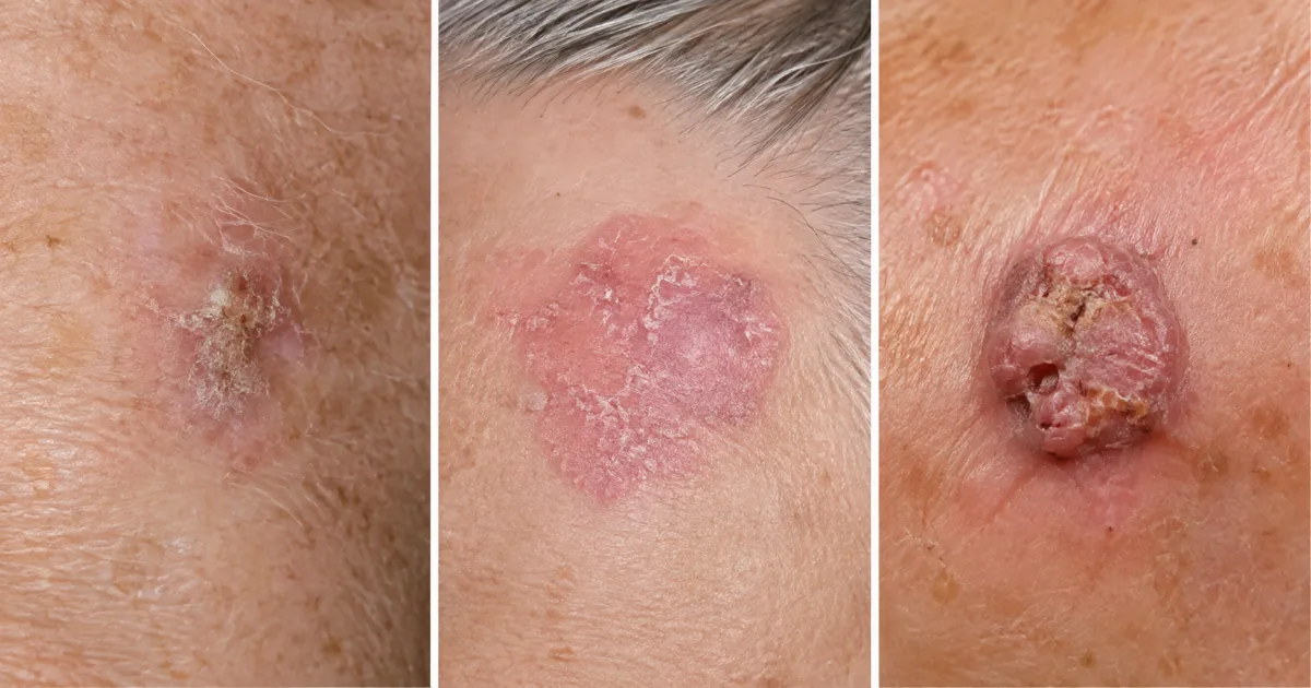
The next step is the carcinoma in situ or Bowen's disease, a cancer that is still confined to the outermost layer of the skin and has not invaded deeper layers. For this reason, its prognosis is excellent and its treatment is straightforward. In a series of 263 carcinoma in situ lesions followed for about eight years, the overall recurrence rate was 6.5%, and it varied depending on the technique used: the lowest rate was observed with surgery. (Övermark et al., 2016)
When tumor cells break through that barrier and begin to invade downward, we are then referring to invasive squamous cell carcinoma. Some forms have their own specific names. The cutaneous horn, for example, is a keratin protrusion that at its base can hide one of these lesions, and the actinic cheilitis it is the equivalent on the lip, an area of special risk. The reassuring idea is that the earlier action is taken along this path, the simpler everything is.
How is it recognized? Symptoms, appearance, and risk areas
Squamous cell carcinoma usually presents as a firm, rough, or crusted lesion that does not heal completely, sometimes painful or bleeding easily. It may look like a persistent sore, a scaly patch, or a growing lump. It appears mostly in areas that have received the most sun exposure over a lifetime. (Firnhaber, 2020)

The highest-risk locations are precisely those most exposed to light, and in some of them, the tumor tends to behave more aggressively. These are the ones I monitor most closely in my practice:
- The lower lip, where it is usually preceded by actinic cheilitis.
- The ear, an area of thin, highly exposed skin.
- The Hairless Scalp, which is common in men with male-pattern baldness.
- The backs of the hands, which accumulates sunlight throughout its lifetime.

None of these signs on their own means you have cancer, but they all warrant a professional evaluation. I'll explain in more detail what to look for in my guide on how to detect skin cancer early. If you want to be sure, you can do so at a online visit or, if you're in Barcelona, in a on-site visit.
How is it diagnosed?
The diagnosis begins with the dermatologist's examination and is confirmed by a biopsy. It is a quick, outpatient procedure that always follows the same sequence:
- Clinical suspicion. The dermatologist assesses the appearance, location, and evolution of the lesion.
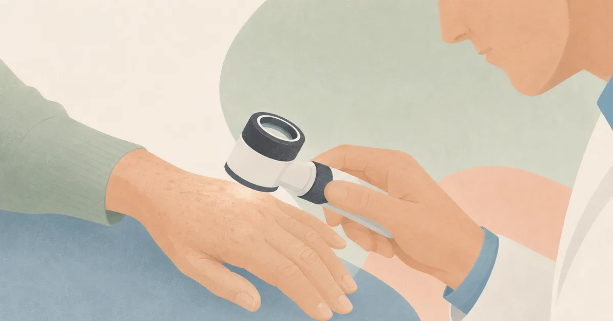
- Dermatoscopy. The same digital dermoscopy The device we use to monitor moles magnifies the skin and reveals structures that are invisible to the naked eye, which helps direct our suspicion even before the biopsy.
- Biopsy. Depending on the appearance of the lesion, a shave biopsy (if it is elevated) or a punch biopsy (from the most abnormal area) is taken and examined under a microscope.
This histological analysis confirms whether it is a squamous cell carcinoma and also provides the information needed to determine the treatment: the degree of cellular differentiation and the depth to which the tumor has spread. In high-risk cases, guidelines recommend supplementing the evaluation with an examination of the lymph nodes in the area and, if necessary, imaging tests. (Firnhaber, 2020) (Stratigos et al., 2020)
What makes a squamous cell carcinoma high risk?
Not all squamous cell carcinomas behave the same way, and the difference depends on a few factors aggression factors which the dermatologist and pathologist evaluate on a case-by-case basis. The more of these factors a tumor has, the greater the risk that it will reappear after treatment (what we call a recurrence) or that it will spread. The vast majority, however, do not have any of these characteristics.
There are four main high-risk factors, and the pathologist always looks for them in the biopsy report:
- 2 cm or larger.
- Poorly differentiated histology: Under a microscope, tumor cells look very different from normal cells.
- Growth toward the deeper layers, underneath the fat.
- An invasion of nerves.
In addition to these, there are three other factors that also play a role: the location in sensitive areas (lip, ear), whether the lesion is ulcerated, and a weakened immune system (immunosuppression). (de Jong et al., 2024)
The best way to see how significant these factors are is through numbers, viewed in context. An international cohort of 16,844 squamous cell carcinomas (from centers in the United States, Spain, and Brazil) analyzed how the prognosis changes depending on how many risk factors the tumor has. Before looking at the table, it’s important to understand one thing: 3 out of every 4 tumors had no risk factors at all, so the majority fall into the first column—the one with the best prognosis. The highest figures on the right correspond to the rare cases that have several risk factors at once. (Ran et al., 2025)
| 5-year outcome | 0 factors | 1 | 2 | 3 | 4 |
|---|---|---|---|---|---|
| Local recurrence | 1,7% | 5,0% | 8,8% | 16,0% | 33,0% |
| Lymph node metastasis | 0,6% | 3,6% | 11,0% | 20,0% | 28,0% |
| Distant metastasis | 0,2% | 1,1% | 2,3% | 7,9% | 8,4% |
| Tumor mortality | 0,3% | 1,9% | 5,4% | 11,0% | 25% |
And this is the point I emphasize most often in my practice: in that majority group with no risk factors, the 5-year tumor-specific mortality rate was just 0.3%. In other words, the gradient in the table does exist, but the vast majority of people fall on the far left—where the prognosis is best—rather than in the columns on the right. (Ran et al., 2025)
Staging systems—that is, methods for measuring the severity of these tumors—are used to classify them. Two of the best-known are those developed by the American Joint Committee on Cancer (AJCC, 8th edition) and Brigham and Women’s Hospital (BWH). A study of 680 head and neck carcinomas compared the two systems and found that the BWH system is more accurate: it identifies cases of true risk with high precision, with a specificity of 93% (that is, it rarely gives a false alarm), and avoids classifying tumors as severe when they are not actually severe. (Ruiz et al, 2019)
Only a professional evaluation can tell you if your injury involves any of these risk factors. If you have doubts, do not wait for your next check-up.
Does it metastasize? What is the prognosis?
The vast majority of squamous cell carcinomas are treated and cured before they spread. Metastasis is possible but rare in common tumors, occurring in less than 4% of cases. If it does occur, it usually affects the regional lymph nodes first and, less frequently, distant organs; it is more common in tumors that exhibit aggressive factors or in people with weakened immune systems.
The main message, therefore, is reassuring: in most cases, the prognosis is excellent, with a 5-year cure rate exceeding 90%. Metastasis is the exception, not the rule, and is concentrated in tumors that exhibit multiple risk factors. (Stratigos et al., 2020)
When the tumor does have the capacity to spread, the first destination is usually the regional lymph node. One fact helps to put into perspective who it really affects, and it is best read with its nuance: it corresponds to transplant patients, a very high-risk population, not the typical squamous cell carcinoma of a healthy person.
In the usual scenario, the probability of metastasis is low and the prognosis is very good. Nerve invasion, when it occurs, is one of the factors that most worsens the prognosis, which explains why the dermatologist carefully looks for it in the biopsy report. (Crawford et al., 2024)
How is it treated depending on the area and the risk?
Squamous cell carcinoma is curable, and surgery is the standard of care. Depending on the tumor’s size, location, and risk level, the dermatologist may choose between surgical excision with margins, Mohs surgery in delicate areas, radiation therapy, or—in advanced disease—immunotherapy.
The first-line treatment for common tumors is surgical excision, which involves removing the tumor along with a margin of healthy tissue and then examining the margins to confirm that it has been completely removed. When the margins are positive, the surgery is extended. It is typically an outpatient procedure with very high cure rates. (Stratigos et al., 2020)
In delicate areas, such as the lip, ear, or around the eye, and in cases of recurrent tumors or those with poorly defined borders, the following comes into play: Mohs surgery, a technique that removes the tumor in layers and examines them under a microscope during the same procedure. This ensures optimal margin control while preserving healthy tissue. In a series of periocular carcinomas treated with Mohs surgery, the recurrence rate was only 3.0% after nearly four years of follow-up. (Weesie et al., 2019)
For carcinoma in situ (Bowen’s disease), which is still superficial, there are more treatment options because the tumor has not invaded deeply. Surgery remains the most effective treatment, but there are alternatives depending on the case, with varying long-term control rates:
- Surgery: recurrence 0.8%
- The most effective treatment option for carcinoma in situ.
- Cryotherapy: recurrence 4.71 TP3T
- Controlled freezing of the lesion, useful in selected cases.
- Photodynamic therapy: recurrence 18%
- Good aesthetic results; offers slightly less protection in the long term.
These figures come from eight-year follow-up data on carcinoma in situ lesions and explain why the decision is always individualized during the consultation: photodynamic therapy It produces excellent cosmetic results and is comfortable, while surgery offers the highest level of control. There is no single "best treatment," but rather the most appropriate one for each injury and each person. (Övermark et al., 2016)
""During the consultation, I always emphasize one point: squamous cell carcinoma, when detected early, is a problem with a simple solution. The question is almost never whether it can be cured, but rather what is the best way to treat it in your specific case.""
When the tumor is locally advanced or has metastasized—which is rare—management is referred to a multidisciplinary committee. Here, a breakthrough in recent years has changed the landscape: immunotherapy. Anti-PD-1 antibodies are now the first-line systemic treatment for advanced squamous cell carcinoma that cannot be treated with surgery or radiation, and cemiplimab was the first to be approved for this indication. (Stratigos et al., 2020)
These drugs work by reactivating the patient's own immune system so that it can recognize and attack the tumor—a mechanism similar to the one I explained in the article on immunotherapy for melanoma. The two available anti-PD-1 agents, cemiplimab and pembrolizumab, showed similar response rates in the trials that led to their approval, in a setting where there had previously been no effective systemic treatment:
The practical conclusion is that even advanced cases, which are the exception, have an effective targeted treatment available today.
Can it be prevented? What follow-up do I need?
Yes, the risk can be reduced, and the pillar of prevention is sustained photoprotection. Cumulative sun damage is the driving force behind squamous cell carcinoma, so protecting oneself from the sun throughout life is what most reduces the appearance of new lesions. The guidelines recommend four specific measures: (Stratigos et al., 2020)
- Reduce exposure during peak radiation hours (12:00 to 16:00).
- Wear protective clothinglong sleeves, wide-brimmed hat, sunglasses.
- Apply sunscreen on a regular basis in areas exposed to sunlight.
- Avoid artificial tanning, a preventable source of cumulative damage.
If you want to learn more about how to choose and apply sunscreen, I wrote a guide on the best sunscreens.
In high-risk patients with multiple injuries, there is also the chemoprevention, which aims to reduce the occurrence of new tumors. The most widely studied option is the nicotinamide, a form of vitamin B3, which in the landmark trial (ONTRAC) demonstrated this reduction compared with a placebo:
There is a compelling reason not to neglect follow-up, and it’s best to read this without getting alarmed.
That is why guidelines recommend regular skin screenings tailored to each individual, with more frequent checkups as the risk increases. This is not a stressful form of monitoring, but rather a routine process designed precisely to ensure that any issues are caught early. If you’ve been treated for squamous cell carcinoma, the realistic outlook for your future is good: with sun protection, simple checkups, and, when appropriate, preventive measures, most of my patients lead completely normal lives.
Basal cell and squamous cell: how do they differ
These are the two most common types of non-melanoma skin cancer and are often confused with one another, but they have important differences. The Basal cell carcinoma is the most common type and almost never metastasizes; the Basal cell carcinoma is the second type, and it does have the ability to spread, even if it is rare. This table summarizes the key differences for the patient.
| Feature | Basal Cell | Squamous cell |
|---|---|---|
| Frequency | The most frequent | The second most common |
| Origin | Basal cells of the epidermis | Keratinocytes; usually resulting from actinic keratosis |
| Metastatic capacity | Exceptional | Possible, though rare (less than 4%) |
| Main risk | Local destruction if abandoned | Dissemination if there are aggressiveness factors |
| General forecast | Excellent | Very good when it is detected early |
The underlying idea is reassuring: both are skin cancers with a very good prognosis when detected early. The practical difference is that squamous cell carcinoma deserves a bit more attention in follow-up, because it can indeed spread in a minority of cases. You can read all about its sibling in the article dedicated to basal cell carcinoma. (Kim et al., 2018)
Frequently Asked Questions About Squamous Cell Carcinoma
Is squamous cell carcinoma dangerous?
Where does squamous cell carcinoma metastasize?
Is squamous cell carcinoma benign or malignant?
What is the difference between basal cell carcinoma and squamous cell carcinoma?
How is squamous cell carcinoma treated?
How does a squamous cell carcinoma start?
Do you have a skin lesion that won't heal or that worries you? An early evaluation is usually simple and almost always reassuring. Choose the option that best suits you.
References
- Kim JYS et al. Guidelines of care for the management of cutaneous squamous cell carcinoma. Journal of the American Academy of Dermatology, 2018. JAAD
- Stratigos AJ et al. European interdisciplinary guideline on invasive squamous cell carcinoma of the skin: Part 1 (epidemiology, diagnosis, prevention). European Journal of Cancer, 2020. European Journal of Cancer
- Stratigos AJ et al. European interdisciplinary guideline on invasive squamous cell carcinoma of the skin: Part 2 (treatment). European Journal of Cancer, 2020. European Journal of Cancer
- Keim U et al. Incidence, mortality and trends of cutaneous squamous cell carcinoma in Germany, the Netherlands, and Scotland. European Journal of Cancer, 2023. European Journal of Cancer
- Feldman SR, Fleischer AB. Progression of actinic keratosis to squamous cell carcinoma. Skin, 2011. PubMed
- Övermark M et al. A Retrospective Study of Treatment of Squamous Cell Carcinoma In Situ. Acta Derm Venereol, 2016. Acta Dermato-Venereologica
- Ran NA et al. Number of Risk Factors and Outcomes in Cutaneous Squamous Cell Carcinoma. JAMA Dermatology, 2025. JAMA Dermatology
- Ruiz ES et al. Performance of the AJCC 8th edition vs the BWH tumor classification system for cutaneous squamous cell carcinoma. JAMA Dermatology, 2019. JAMA Dermatology
- de Jong E et al. Cumulative incidence and risk factors for cutaneous squamous cell carcinoma metastases in organ transplant recipients (SCOPE-ITSCC). Journal of the American Academy of Dermatology, 2024. JAAD
- Collins L et al. Skin Cancer and Immunosuppression. Dermatologic Clinics, 2018. PubMed
- Fox M et al. Nodal staging of high-risk cutaneous squamous cell carcinoma. Journal of the American Academy of Dermatology, 2018. JAAD
- Crawford L et al. Perineural spread of cutaneous squamous cell carcinoma via the ophthalmic nerve. Head Neck, 2024. PubMed
- Firnhaber JM. Diagnosis and Treatment of Basal Cell and Squamous Cell Carcinoma. American Family Physician, 2020. American Family Physician
- Weesie F et al. Recurrence of periocular keratinocyte carcinoma after Mohs micrographic surgery. Br J Dermatol, 2019. PubMed
- Migden MR et al. PD-1 Blockade with Cemiplimab in Advanced Cutaneous Squamous-Cell Carcinoma. New England Journal of Medicine, 2018. New England Journal of Medicine
- Hughes BGM et al. Pembrolizumab for locally advanced and recurrent/metastatic cutaneous squamous cell carcinoma (KEYNOTE-629). Ann Oncol, 2021. Annals of Oncology
- Chen AC et al. A Phase 3 Randomized Trial of Nicotinamide for Skin-Cancer Chemoprevention (ONTRAC). New England Journal of Medicine, 2015. New England Journal of Medicine
- Hardin J, Mydlarski PR. Systemic retinoids: chemoprevention of skin cancer in transplant recipients. Skin Therapy Lett, 2010. PubMed