







Melanonychia, that dark line that appears on a nail, is one of the reasons for consultation that generates most concern among my patients. The good news is that the vast majority of these pigmented bands are benign. Subungual melanoma accounts for only 1.9% of all melanomas.. [(Tsiogka et al., 2024)].
Some time ago, I gave a talk to a group of patients about moles and melanoma on the nails. Seeing the interest and concern on their faces, I realized the importance of spreading this knowledge to all my patients and readers.
My name is Sebastian Podlipnik and I am Dermatologist at the Melanoma and Skin Cancer Unit of Hospital Clínic de Barcelona. During my years of experience I have evaluated many patients with melanonychia and I can tell you that knowing how to differentiate when a line on the nail requires attention and when you can rest easy is critical.
What is melanonychia and why do dark lines appear on the nails?
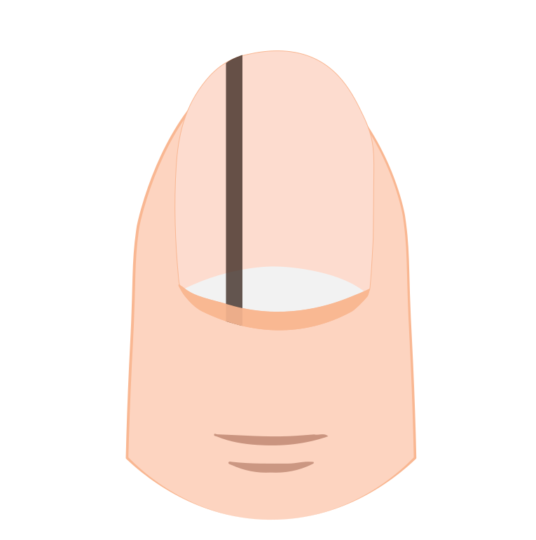
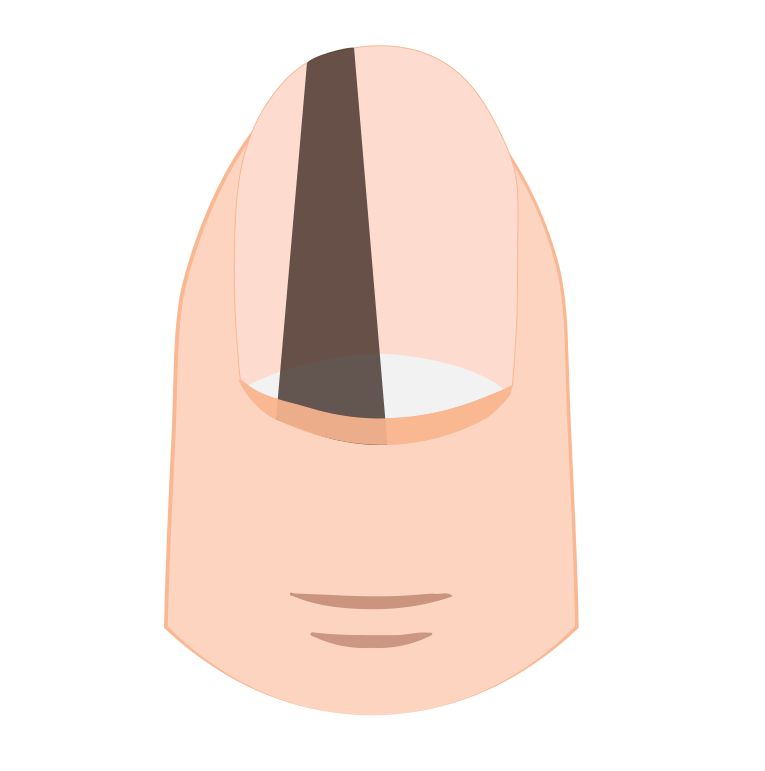
Melanonychia is a clinical sign, not a disease. It refers to the presence of a brown or black pigmented band running longitudinally across the nail. When this band runs from the base to the free edge of the nail, it is called longitudinal melanonychia.
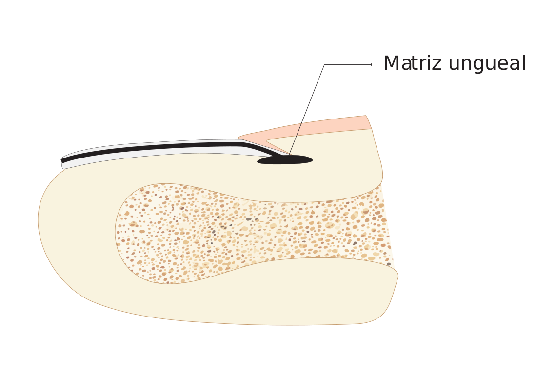
The cells that produce the pigment (melanocytes) are found in the nail matrix, the most proximal area from which the nail plate grows. When these melanocytes are activated or proliferate, the pigment adheres to the nail and moves towards the free edge as the nail grows. It is like a conveyor belt that leaves a continuous mark.
- Longitudinal melanonychia
- Pigmented band that runs longitudinally along the nail, from the base to the free edge. It is not a diagnosis by itself, but a clinical sign that may correspond to benign causes (the majority) or, in a small percentage, to melanoma.
What do moles look like on the nails?
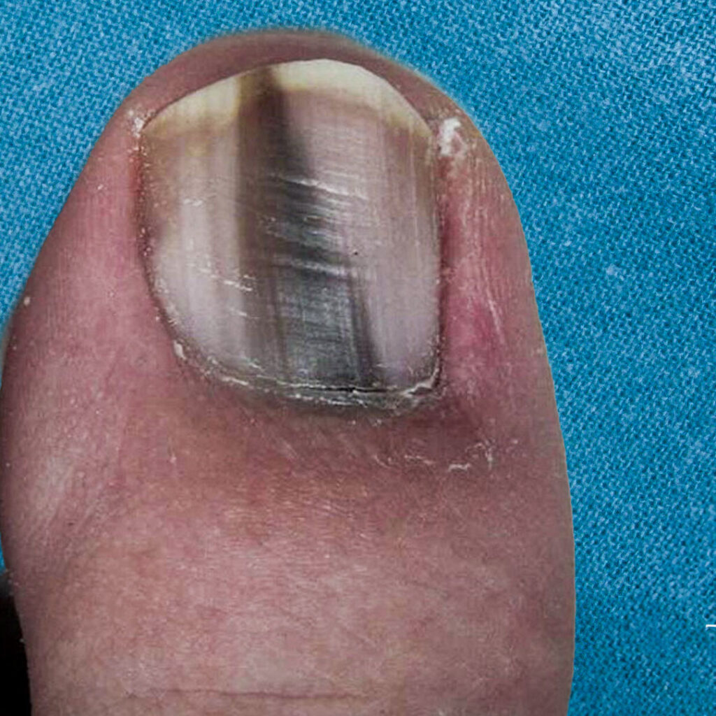
Nail moles (nail nevi) are very stable lesions that do not change significantly over time. They appear as a regular, usually narrow, well-demarcated, homogeneously colored band of pigment affecting a single nail.
With dermoscopy (a special microscope used by dermatologists), moles show a pattern of parallel lines regular in color and thickness. This ordered pattern is the key that allows us to differentiate a benign mole from a suspicious lesion. Regular parallel lines on dermatoscopy indicate benignity..
What signs should lead one to suspect melanoma of the nail?
Nail melanoma differs from benign moles in that it presents progressive changes, irregularity and extension of the pigment beyond the nail. Although it is a rare form of skin cancer, If it is detected early, early detection is essential because the prognosis is greatly improved with early diagnosis.
ABCDEF rule for nail melanoma
Dermatologists use the ABCDEF rule as a guide to evaluate pigmented nail bands. [(Levit et al., 2000)].
- A (Age): Nail melanoma is more frequent between the ages of 50 and 70, and in people of high phototypes (African Americans, Asians, Hispanics).
- B (Band): Brown or black band more than 3 mm wide with irregular or fuzzy edges
- C (Change): Any rapid change in color, size or shape of the band
- D (Digit): The thumb and the first toe are most frequently affected
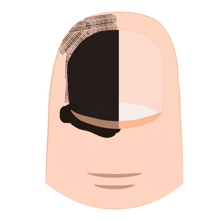
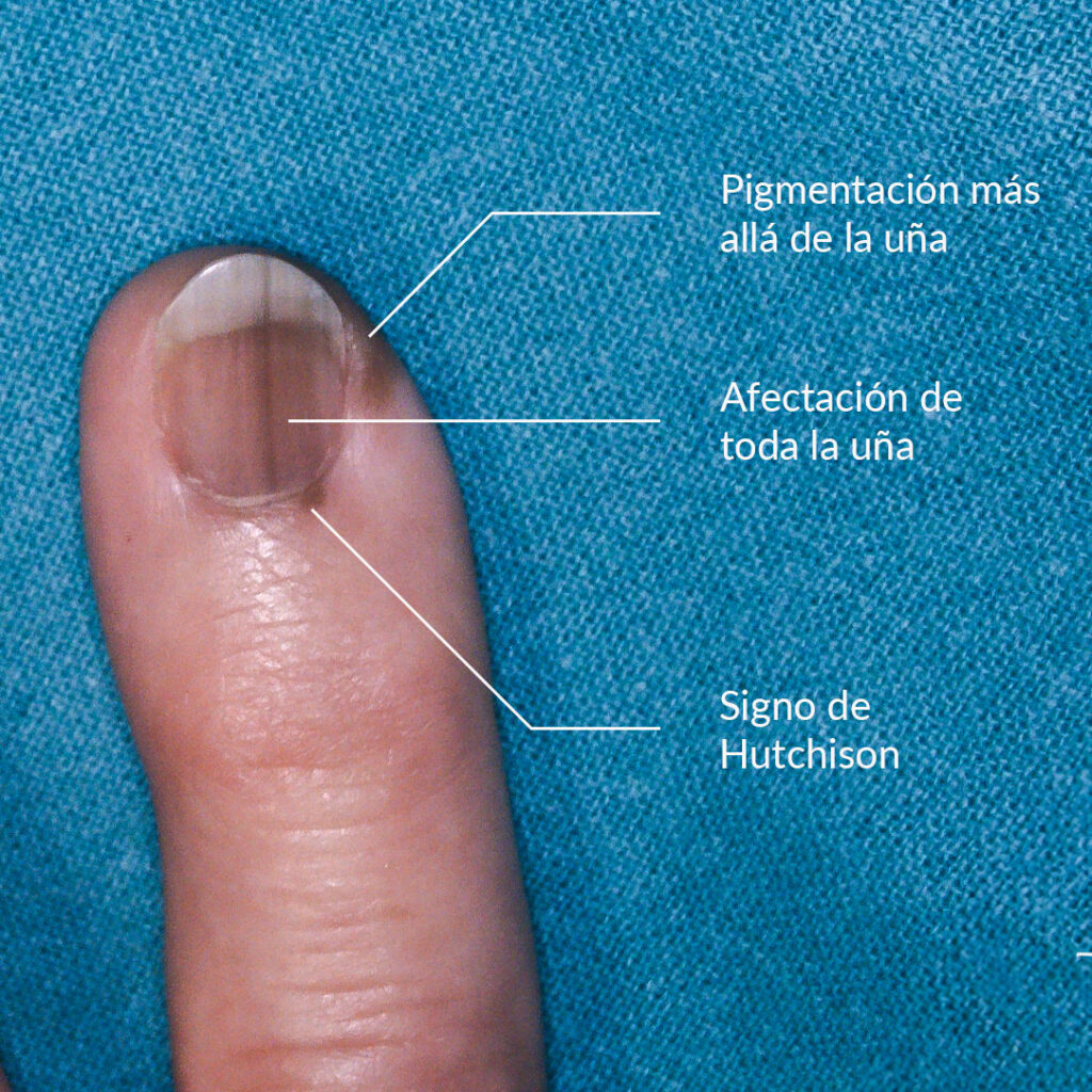
- E (Extension): Pigmentation extending to the nail fold or periungual skin (Hutchinson's sign)
- F (Family): Personal or family history of melanoma
A study that evaluated 84 melanonychia biopsies found that a band greater than 40% of the nail width is the best predictor of melanoma. (p=0,002). [(Ko et al., 2019)]
Progressive changes
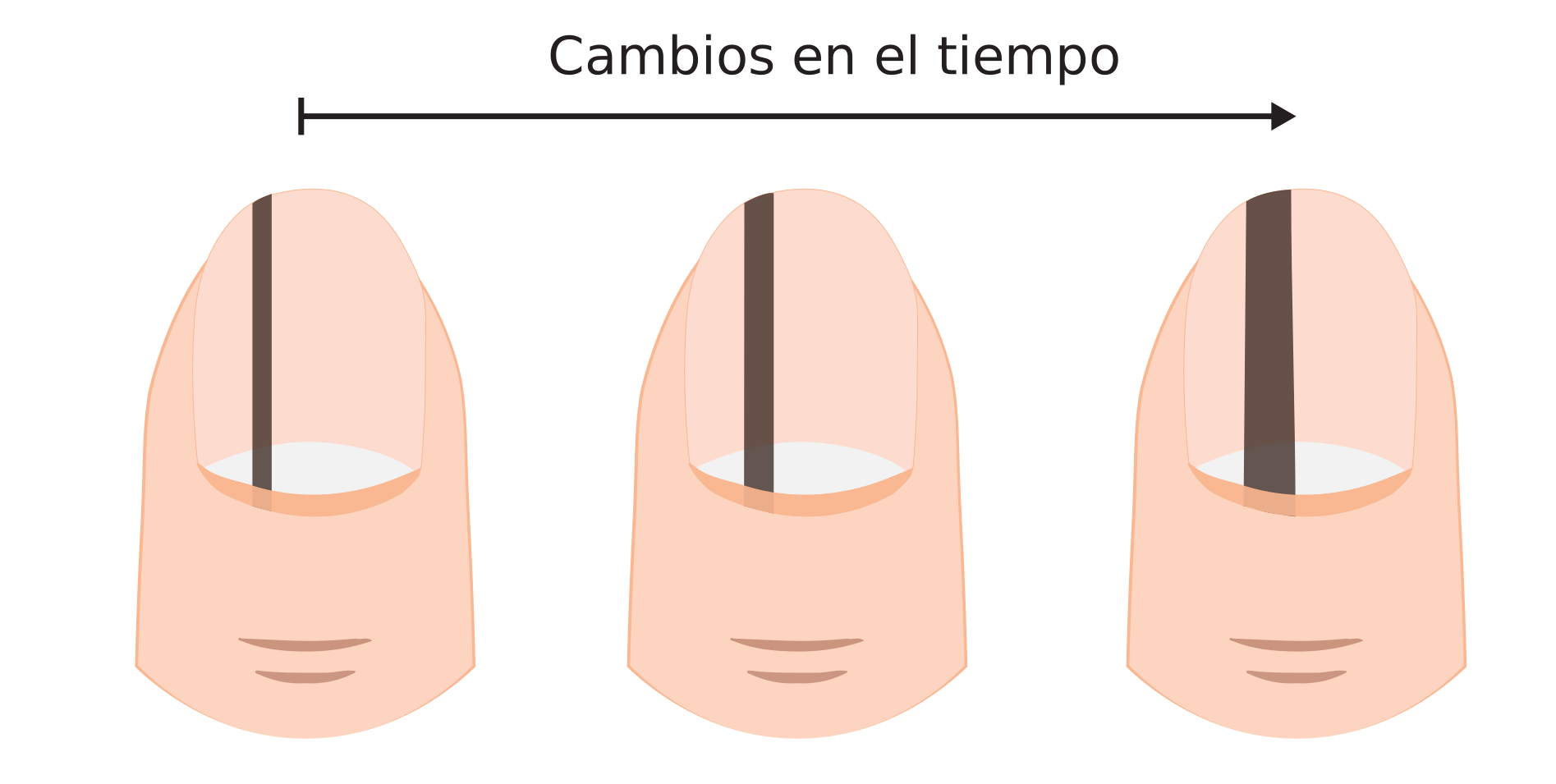
Benign nail moles are very stable. One of the first signs of melanoma is progressive change: the band becomes wider, darker or changes shape within weeks or months. Nail melanoma grows, benign mole remains stable.
Triangular shape of the pigmented band
If a lesion grows rapidly, the newer pigment (near the matrix) will be wider than the older pigment (at the free edge). This generates a characteristic triangular shape which is an important warning sign.
Other important signs
- Greater than ⅓ thickness of the nail: If the pigment band occupies more than one-third of the total width of the nail
- Pigmentation beyond the nail: When the brown pigmentation spreads to the skin surrounding the nail
- Hutchinson's sign: Pigmentation affecting the proximal nail fold, one of the most specific signs of nail melanoma.
- Destruction of the nail plate: When the nail is broken, deformed or lifted up
Consult your dermatologist if you notice
- A new dark band on a single nail that you had not noticed before
- A progressive change in the width, color, or shape of an existing band
- Pigmentation spreading to the skin around the nail
- Destruction or deformation of the nail plate together with pigmentation.
Benign vs. malignant melanonychia: how to tell the difference?
Most melanonychia are benign. A dermoscopic study found that the most useful signs to suspect malignancy are band asymmetry (OR 34.0), Hutchinson's sign (OR 18.2) and the presence of multiple colors (OR 11.6). Band asymmetry is the strongest predictor of nail melanoma. [(Ohn et al., 2018)]
| Feature | Benign melanonychia | Suspected melanoma |
|---|---|---|
| Bandwidth | Narrow (<3 mm) | Wide (>3 mm or >40% of width) |
| Edges | Regular and well-defined | Irregular and diffuse |
| Color | Homogeneous (single tone) | Heterogeneous (multiple tones) |
| Evolution | Stable over time | Progressive changes |
| Hutchinson's sign | Absent | Present |
| Number of nails | It may affect one or more | Almost always a single nail |
He recalls a 62-year-old patient who came for consultation because his wife had noticed that the dark line on his thumb was widening. He had had it for years and had never given it any thought. Dermoscopy showed an irregular pattern and it eventually turned out to be melanoma in situ. Detection was timely and treatment was conservative, with no need to amputate the finger.
"Most dark lines on the nails are benign. But if you notice a change, don't let it go. Consulting early can make all the difference."
Dr. Sebastian Podlipnik
Do you have a spot on your nail that worries you?
I can evaluate it with dermoscopy and give you an accurate diagnosis.
Melanonychia in children: should I be concerned?
In children, longitudinal melanonychia is almost always benign. A meta-analysis of 1,391 pediatric patients found that 86.3% corresponded to benign nevi. No invasive melanoma has been reported in children with melanonychia.. [(Tsai et al., 2024)].
This is very reassuring for parents who come to my office concerned about a dark line on their child's nail. In the vast majority of cases, the correct approach is to perform periodic follow-up with digital dermoscopy, without the need for biopsies or surgery.
One detail that I always explain to parents: melanonychia in children can change appearance during growth and this does not necessarily indicate malignancy. The important thing is that these changes are evaluated by a dermatologist with experience in nail lesions.
Are there other causes of dark nail lines?
Many conditions can produce pigmented bands on the nails in addition to moles and melanoma. Broadly speaking, the causes fall into two groups: melanocytic activation (melanocytes produce more pigment than normal) and melanocytic proliferation (the number of melanocytes increases, as in moles and melanoma).
A key difference is that melanocytic activation usually affects several nails at once, whereas moles and melanoma typically affect a single nail. If several nails are affected, the cause is usually benign..
Drug-induced melanonychia
Several medications can cause melanonychia as a side effect. The most frequent are:
- Chemotherapy: Cyclophosphamide, doxorubicin, 5-fluorouracil
- Antiretrovirals: Zidovudine (AZT), the most known to cause melanonychia.
- Antimalarials: Chloroquine, hydroxychloroquine
- Others: Minocycline, psoralens (used in phototherapy)
Melanonychia in dark skins
In people with high phototypes (dark skin), melanonychia is frequent, physiological and does not require treatment. It can affect multiple nails and does not require any treatment. However, it is important to have a dermatologist confirm that it is racial melanonychia and not another cause.
There are also other common causes of melanocytic activation such as pregnancy, repeated trauma (nail biting, narrow shoes), infections, lichen planus, psoriasis, Addison's disease and hyperthyroidism.
What do we do in the dermatological practice when we see a melanonychia?
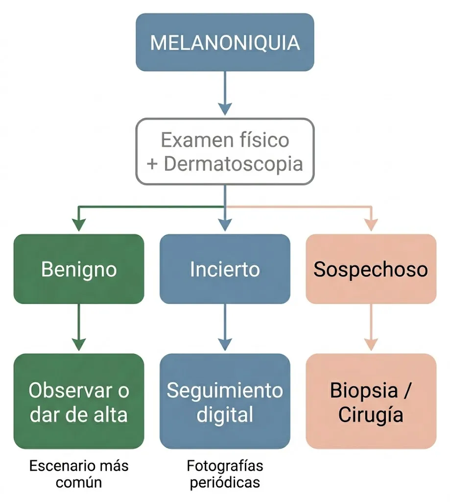
To properly evaluate a melanonychia, we follow a systematic three-step process that allows us to make the best decision for each patient. Excisional biopsy of the nail matrix remains the gold standard for definitive diagnosis, but not all melanonychia require biopsy. [(Ricardo et al., 2025)].
1. Physical examination
The first thing I do is a complete physical examination of all your nails, both fingernails and toenails. I evaluate for deformity, whether the melanonychia affects one or more nails, and analyze the color, shape and size of the band. In addition, I review your medical history to identify conditions or medications that may explain the pigmentation.
2. Dermatoscopy (onychoscopy)
I then use the dermatoscopy, I use a special microscope that allows me to examine the band pattern in great detail. The most useful dermatoscopic signs to suspect malignancy are asymmetry, the presence of multiple colors and irregularity of the lines. [(Ohn et al., 2018)]
One piece of data that reflects the complexity of this diagnosis: a study of 152 dermatologists found that the overall accuracy for diagnosing nail melanoma was 46-55%, regardless of experience level. [(Di Chiacchio et al., 2010)]. This reinforces the importance of combining clinical evaluation with dermoscopy and, when in doubt, digital follow-up or biopsy.
3. Take a behavior
After clinical and dermatoscopic evaluation, there are three possible scenarios:
- Clearly benign lesion: We do nothing or discharge. This is the most frequent scenario
- Doubtful injury: We perform digital follow-up with periodic photos to detect changes over time. We can also request a partial biopsy (nail clipping) to get more information.
- Suspicious lesion of malignancy: We directly propose an excisional biopsy or surgery to remove and analyze the entire lesion.
Treatment of nail melanoma: amputation is no longer always performed
The treatment of nail melanoma has changed greatly in recent years. Surgical management has evolved from aggressive amputations to conservative approaches that preserve the digit. [(Zhang et al., 2021)].
For in situ (confined to the epidermis) or minimally invasive melanomas, the current trend is for conservative functional surgery, which removes the lesion while preserving the toe. Amputation is reserved only for cases with bone invasion or advanced disease. [(Darmawan et al., 2022)].
But most melanonychias do not require surgery. If after the evaluation we conclude that the lesion is benign, the attitude is simply to observe or perform a periodic follow-up. Only when there is suspicion of malignancy do we consider surgical treatment.
Early diagnosis is what makes the difference in prognosis. A melanoma detected in situ has an excellent prognosis and can be treated conservatively. If you want to know more about how it is classified, I explain it in my article on the prognosis of melanoma.
Frequently asked questions about melanonychia
Is melanonychia cancer?
No. Melanonychia is a clinical sign that describes the presence of a pigmented band on the nail. The vast majority of melanonychia are benign and are due to moles, melanocytic activation by drugs, trauma or racial factors. Only a very small percentage corresponds to melanoma (1.9% of all cutaneous melanomas).
Is it normal to have a black line on the nail?
It can be completely normal, especially in people with dark skin, where longitudinal melanonychia is physiological and frequent. It may also be due to minor trauma or certain medications. However, if it is new, affects a single nail or is changing, it should be evaluated by a dermatologist.
When should I go to a dermatologist for a spot on the nail?
You should consult if the band is new and you have not noticed it before, if it is changing (widening, darkening or changing shape), if the pigment extends to the skin around the nail, or if the nail is deformed. If you have any doubts, it is always best to ask for advice to put your mind at ease.
Can nail melanoma be cured?
Yes, especially when detected in early stages. Melanoma in situ has an excellent prognosis and can be treated with conservative surgery, without the need to amputate the finger. That is why I insist on the importance of not delaying consultation in the event of any suspicious change.
Is melanonychia in children dangerous?
In the vast majority of cases, no. A meta-analysis with 1,391 children found that 86.3% were benign nevi and no invasive melanoma was reported in pediatric age. Even so, it is recommended that a dermatologist evaluate the lesion and perform periodic follow-up.
Consultation with a melanoma specialist
If you have a pigmented nail band that concerns you, a dermatology consultation can give you peace of mind or, if necessary, an appropriate follow-up plan. Most of the time you'll leave the consultation knowing that everything is fine. And if not, you will have taken the most important step: detect in time. If you want to learn more about how to identify malignant moles or know the types of skin cancer, I invite you to read my guides. You may also be interested in learning more about the moles on soles of feet and palms, another special localization of acral melanoma.
Do you want me to evaluate your melanonychia?
I can help you determine if you need follow-up or treatment.





55 Responses
Hello Doctor, I had a very thin line at the end of my nail or finger. One day I accidentally inserted a staple remover clip just a little bit. The staple remover clip was actually wider than the line, haha, and when the clip passed over the line, it took away little pieces. I was able to remove as much as I could. Now I just have to wait for the nail to grow out and for it to disappear completely. My question is, does nail melanoma affect the skin and nail, right? Meaning, would I have been able to remove that line with a clip, or not?
Hi Samantha, it's difficult to determine the cause just from what you've told me. It's best for a.
Should I be alarmed if the cause is something other than what you're saying?
Hello, I had a line on my fingernail and I measured it. It was on my thumb, 6 mm long. It appeared in the middle of my nail. It was very thin. I even thought it was made with a pen (it wasn't like the pictures shown, just a line on the nail). I didn't realize it at first. I saw this article and it interested me. The line gradually disappears as my nail grows. It didn't start from the cuticle, if that makes sense? I saw it in the middle of my nail, it wasn't long enough to cover from the beginning to the end. It's disappearing now. I see that some causes are X-ray treatments. A few weeks or days before I noticed this line, I had X-rays and a CT scan of my chest. Is this alarming? Right now, the size of this line is, I think, a pen mark, almost entirely gone because I cut my nail.
Hello Adrian, surely from what you're saying and that it's disappearing, it's nothing serious. The best thing is to keep an eye on it and if it all disappears, I would be at ease.
Hello, a very thin line appeared on my right thumb. The line was about 4 or 5 mm long and about the width of a hair, meaning very thin. This line did not appear on my cuticle, so as the nail grows and I cut it, the line starts to disappear. Right now, the width is still the thickness of a hair, and its length is approximately 1 mm because it is going away. Is this something to be concerned about?
Hello Erasto, if the line appears to be disappearing, it's not something you should worry about, as it's likely a benign condition. Nails can be complex to diagnose without an in-person examination.
Hello! I have been undergoing treatment for over 5 years with Metoject (injected MTX). I have had a black line on one of my fingernails for at least a year. Could the cause of this melanocytic activation possibly be Raynaud's syndrome or treatment with Methotrexate? I am not worried because thanks to your article, I see that it seems to be just melanocyte activation. It does change as the nail grows, and I haven't hit it.
In addition, I am in perimenopause, and I have seen on gynecology websites that moles can change due to hormonal changes. I'm mentioning this because I've also had several grow on my back. And maybe this could also be the reason for the nail problem... right? Thank you very much. I really liked the article because it's so easy for everyone to understand.
Hi Natalia, it's possible that Methotrexate or Raynaud's syndrome are related, but it's important for a dermatologist to examine your nail to rule out any other issues. An in-person evaluation is best in this case.
Hello,
I read your article and I'm a little worried. Last year, a line appeared on my thumb. I went to a dermatologist and an oncologist, and after examining me with a small microscope, they both agreed it was a mole. However, a year has passed since then, and another line has now appeared on the same nail, but it hasn't shown any dark coloration around the edges. Do moles grow?
Hello Stephanie, given that the line has reappeared, it would be good to have a dermatologist check it again. Although there are no alarming signs, a consultation will give you greater peace of mind.
Hello, I have a very thin black line on my thumb, the thickness of a hair. However, as I trim my nail, it recedes. It advances with the nail, and today it's almost gone. Should I be concerned?
Hello Eduardo, a dark line on the nail can be due to several causes, from mild trauma to other conditions. It's positive that the line is disappearing, but I would recommend visiting a dermatologist for a more detailed check-up to ensure it's nothing serious.
Could a blow or trauma to the nail from footwear or hitting it with a metal point cause a dark line on the nail? I have worn safety boots a lot and I have had injuries to my nails. One is a light brown dark line, and another of the same color but on the sides. I want to assume they are injuries, but I really don't know. I clarify that they have not grown or presented abrupt changes over time.
Hello Raúl, dark lines on the nails can be caused by trauma, nail dystrophies from repeated trauma, but there are also other possible reasons. Since you've noticed these changes and haven't had any hard impacts, it would be advisable for a dermatologist to examine your nails to rule out any other conditions.
Hello, I read your article and found it interesting, as I have a spot similar to those in the photos on my toe. By any chance, do you or any colleague of yours offer consultations in Mexico?
Hello Sylvanna, if you have a spot similar to those you've seen in photos, it would be best to have a dermatologist examine it. This will allow for a proper evaluation and, if necessary, the corresponding treatment. We do not provide services in Mexico, but I suggest you look for a local specialist.
Hola buenas, tego una duda que si esto se puede contagiar porque a varios de mis familiares les ha salido una línea oscura en alguno de sus dedos
La melanoniquia, o la aparición de una línea oscura en las uñas, puede deberse a varias causas, como:
Melanoniquia fisiológica: Común en personas con piel más oscura, es una condición normal y no contagiosa.
Trauma o lesión: Golpes o presión constante pueden causar líneas oscuras.
Infecciones: Fúngicas o bacterianas, aunque rara vez son contagiosas.
Condiciones médicas: Trastornos sistémicos pueden manifestarse en las uñas.
Melanoma: Un tipo raro de cáncer que necesita evaluación médica urgente.
La mayoría de las causas no son contagiosas. Se recomienda consultar a un dermatólogo para un diagnóstico preciso, especialmente si la línea es nueva o cambia.
Hola, recientemente me empecé a alterar un poco porque supe de la existencia de los melanomas, yo he tenido una linea marrón muy clarito en mi uña del pulgar derecho de la mano desde hace muchos años, yo tengo 23 años y jamás se ha modificado. Sin embargo todavía me genera inquietud, podría ser solo un lunar?
Jamás le di importancia la verdad pero ahora sí me inquieta un poco, nuevamente comento que jamás ha crecido esa linea y es uniforme
Hola, Ángel. Una línea marrón en la uña que no ha cambiado en años y es uniforme lo más probable es que sea un lunar, especialmente si no presenta otros síntomas como crecimiento o cambios de color. Sin embargo, es importante que consultes con un dermatólogo y obtener una evaluación médica para estar tranquilo.
Hola, soy Manuel. En noviembre de 2023 detecte una línea oscura pero tenue en la uña más pequeña del pie izquierdo. Al principio pensé se debía a un golpe o algo así pero ahora estamos en julio de 2024 y esa línea sigue igual, no se ha hecho ni más grande ni más oscura, sigue igual solo que ahora noto que surgió otra línea en la misma uña solo que menos oscura y con una separación entre ellas de aprox 3 o 4 mm. No veo manchas oscuras en la piel como señala en su publicación pero si me preocupa que no desaparezcan y que ahora sean dos. Gracias.
Hola, Manuel. Una línea oscura en la uña que no cambia de tamaño o color con el tiempo suele ser menos preocupante. Sin embargo, ya que ha notado una nueva línea y un cambio en la separación, sería prudente que consultes con un dermatólogo. Es importante que un profesional evalúe cualquier cambio en las uñas para descartar condiciones serias.
Hello, good day. My 7-year-old niece started with a thin line on her index finger, and it has now grown to cover the entire width of the nail in about 1 year. Initially, she was seen by a doctor, and they indicated to monitor its growth. Now, she has had this sudden growth, and the proximal nail fold also looks a bit darker. She already has an appointment scheduled. Could this be concerning? Thank you!
Hi Alex, malignant nail lesions in children are extremely rare. It is likely nothing serious, but if the changes persist or worsen, it would be advisable for a dermatologist to examine her to rule out any other problems.
My son has a "mole" on his left thumb, we are doing studies to rule out melanoma, how much does an operation to remove it cost?
Hi Mariel, first of all, if the dermatologist establishes that it is a benign mole you will not have to do any surgery. If your son is a boy, it will almost certainly be a benign mole.
Hi I am prepared I have a very thin line about 2 mm on my index nail and there is a little dot in the flesh above the nail should I be worried?
Hi Andres, I think there is nothing to worry about but you should go to a dermatologist to have it checked. Best regards
Is there a possibility that longitudinal melanonychia may go away on its own over time?
This doubt arises because if it comes out of the blue (caused by a possible trauma), can the resolution of it be given the same if we consider that it was caused by a trauma?
Hi Sergio, exactly, if the mechanism of melanonychia production is traumatic it is possible that it will go away with time.
Best regards
Hello I am a little worried but it is not a very dark black spot, what if I was sent several nails with lines that look like spots but I want to know what you tell me.
Hello, I understand your concern. If the lines appear on several nails at the same time, it is most likely a benign condition. However, it is always best to have a medical evaluation to be sure.
I have had a black nail since I was a little boy with a streak that does not grow and now suddenly all my nails have stains should I be worried?
Hello Arfhad, Most likely it is nothing important as there are several nails affected. However, if you are concerned it is always advisable to see a dermatologist for a checkup.
Best regards
Hi, I am 16 years old and recently I got straight black lines on more than one nail, looking at your report according to me they are moles but still I am very worried.
Hello, probably because of your age and the presence of multiple nails, it is nothing important. However, it is never too much to go to the dermatologist for a personalized assessment. Best regards
Hi, I have a spot under the nail of my big toe. It's not a line, it's a spot and I didn't hit it or anything, it just appeared out of nowhere. Should I be worried? I was looking for information because I started with an intense pain under the nail of the big toe of my left hand, I have a thin white line that does not go away, but it has never hurt like today. What can you tell me about it? Thank you
Dear Pame, I would love to help you online but to properly evaluate the nails you need to see them in person.
Good morning. A year ago I noticed that I had a very thin black line and I thought it might be a splinter, I cut the nail but it is still there. The nail on my left hand is always deformed and I lose it, but for some time now this line has grown, and now I have another line on my right hand nail, thinner at the top and wider at the bottom.
Hello! I think it is important to have it evaluated by a dermatologist to be sure that it is nothing important. Best regards
hello i have had a scratch on my thumb nail for 10 years and it hasn't changed or grown should i be concerned?
Hi Luna. If nothing has changed in 10 years it is very unlikely that it is something important. Although the recommendation is always to go to your dermatologist to check it with a dermatoscope to make sure there is nothing suspicious.
Hello, I got a black dot under my nail. I cut it out to rule out a splinter, but no. It is in the flesh of the nail. It's in the flesh of the nail and it's like a little mole. Oh, it's under the little fingernail. And I have lupus, I don't know if it's a product of it.
Hello Lola, it is possibly a small splinter hemorrhage. If it does not disappear in 3 months it is best to consult a dermatologist.
Thank you
I have moles on my nails, on all my fingernails, and on some toenails, although they have been disappearing, I know they are not malignant because they have not affected me in any way, they even grow well without any problem. Should I be worried?
Dear Stiven, if you have spots on several nails at the same time it is very unlikely to be a malignant lesion.
Hi Sebastián, well, I have them on almost all my nails (toes and hands), some lines are very thin, on others they join more. But they've never formed a triangle like you say in the article, nor has the skin around them turned black. But still, I'm always a little scared. I don't know what to do. I prefer to think they're moles, and as you say, since there are several spots at the same time, I doubt it's some kind of symptom, right? I went to the doctor once, but they didn't even know what was wrong with my nails (I live in Colombia).
Two months ago I noticed that I had a line on the nail of my right index finger, it has been growing, not so much, but it is wider now, I have been examined by 3 dermatologists but they tell me that I should not worry, but I am worried that I will not have a diagnosis in time.
Hello Aldair. In your case it would be necessary to evaluate this spot live and certainly follow it over time to see how it evolves.
And if the line I have is less than 2 ML and has not grown, what is the reason?
?
Hi William, First of all it is important to evaluate it with dermatoscopy to see what it is. If it is less than 2 mm and does not change or grow, it is most likely due to a mole on the nail. But it is always better to confirm it with the expertise of a dermatologist.
I was worried about a mole I've had since I was a little girl but I'm much calmer reading this information Thank you!
Thank you very much for your comment Eva!